| Theories and Techniques of Oral Implantology (vol.2) (published 1970) | Dr. Leonard I. Linkow |
|
|
Next Page |
| This is an archival HTML version of this book originally hosted here in 2006. The HTML may not display well on modern browsers. Please view the modern PDF Version for a better viewing experience. |
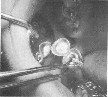
Fig. 15-65. The superstructure was then cemented over the built-up acrylic core and gold post extending from the template.
Atypical implant situations 649
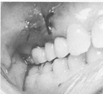
Fig. 15-66. The occlusion was once again checked. Sutures were used to close the wound.
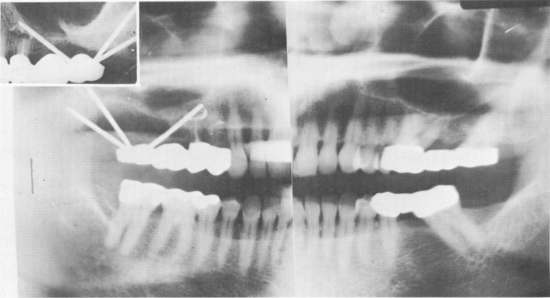
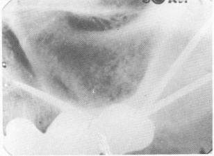
Fig. 15-67. A Panorex and intraoral radiograph showing the completed case. (From Linkow, L. I.: Atypical implantations for anatomically contraindicated situations, Dent. Concepts, Fall, 1967.)
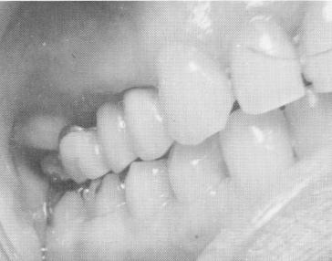
Fig. 15-68. Three weeks later, the tissue looks excellent.
Fig. 15-69. An 18-month postoperative x-ray shows filling-in of bone and the implant in excellent condition.
|
|
Page 649 |
Next Page |
|
Copyright warning: This information is presented here for free for anyone to study online. We own exclusive internet copyrights on all content presented on this website. We use sophisticated technology to identify and legally close down websites that reproduce copyrighted content without permission - so please don’t do it.
|