| Theories and Techniques of Oral Implantology (vol.2) (published 1970) | Dr. Leonard I. Linkow |
|
|
Next Page |
| This is an archival HTML version of this book originally hosted here in 2006. The HTML may not display well on modern browsers. Please view the modern PDF Version for a better viewing experience. |
Atypical implant situations 639
A
C
B
D
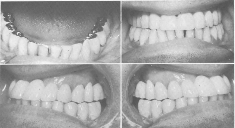
Fig. 15-30. A, The two mandibular unilateral partial dentures are cemented into position with hard cement. B to D, The completed case shows the elimination of the Class III malocclusion.
Fig. 15-31. A severe cross bite relationship.
both jaws at rest. This does not necessarily mean in-creasing the vertical dimension. An illustrative case is shown beginning with Fig. 15-31. The patient had previously undergone bite-raising procedures that opened his bite with removable appliances but that caused great discomfort and pain in his temporomandibular joints. His preoperative Panorex revealed very few maxillary teeth (Fig. 15-32) .
Using the patient's removable denture as a plat-form, the operator molded a thick mix of acrylic over the denture and over the existing prepared teeth while the patient's jaw was in the most retruded position. It was imperative to maintain as close a bite as possible during this procedure in order not to increase the patient's true vertical dimension. Thus the bite was opened just enough to allow the molded max-
illary acrylic teeth to come edge to edge with the mandibular incisors. Once this had been accomplished, the temporary teeth were trimmed and polished. The physiologic rest position was immediately checked. If no rest position existed, it would have been necessary to shorten the acrylic teeth.
The patient wore the provisional splint for about 4 weeks, with no pain or discomfort. Using the "built-up" acrylic teeth on the removable denture as a bite guide, the operator fabricated impressions and castings for the teeth on the right side of the arch (Fig. 15-33). Blade implants were then placed on both sides of the maxillary arch (Fig. 15-34). All remaining mandibular teeth were prepared, and blade implants were placed in both posterior endentulous areas (Fig. 15-35).
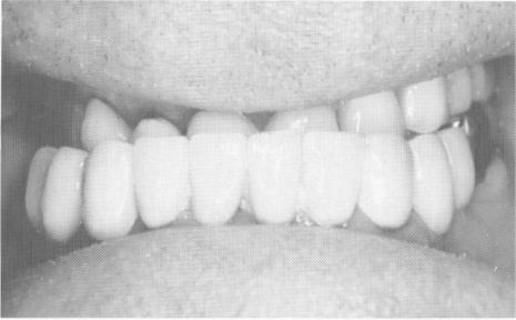
A full arch fixed porcelain-baked-to-metal pros-thesis was completed for each arch, placing the jaws in a Class I relationship (Fig. 15-36) . A Panorex of the completed case shows the blade implant supports (Fig. 15-37).
Case 4
Restoring centric occlusion by occlusal reconstruction and a blade-vent
Another case of extremely poor occlusion re-stored with the help of an endosseous implant is that of a 46-year-old male patient. The patient, because of a traumatic injury during his childhood, was missing half of his left condyle. His maxillary
|
|
Page 639 |
Next Page |
|
Copyright warning: This information is presented here for free for anyone to study online. We own exclusive internet copyrights on all content presented on this website. We use sophisticated technology to identify and legally close down websites that reproduce copyrighted content without permission - so please don’t do it.
|