| Theories and Techniques of Oral Implantology (vol.2) (published 1970) | Dr. Leonard I. Linkow |
|
|
Next Page |
| This is an archival HTML version of this book originally hosted here in 2006. The HTML may not display well on modern browsers. Please view the modern PDF Version for a better viewing experience. |
612 Theories and techniques of oral implantology
A
B
C
D
E
F
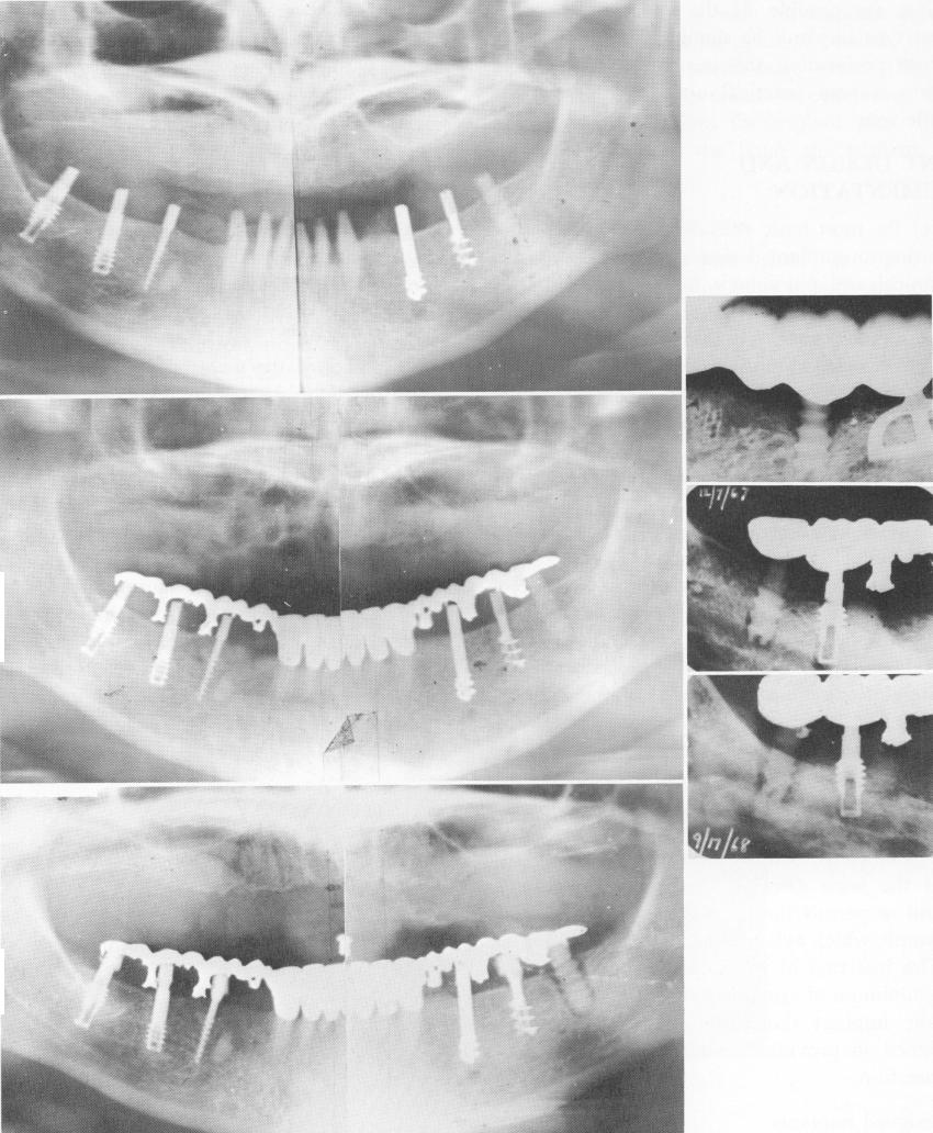
Fig. 14-6. A, Six different implants are seen in the same mouth. Left to right: Two Linkow titanium vent-plants, a narrow ridge (M. Chercheve) implant, a Chercheve spiral-shaft implant, a Muratori spiral implant, and a Sandhaus crystalline bone screw (aluminum oxide). The picture was taken immediately after the implants were placed. B, Three months later a breakdown of bone had started around the synthetic sapphire implant. C, Fourteen months postoperatively, the continued breakdown around the crystalline bone screw, but not around any of the others, is clearly shown. D, A blade-vent is acting as anchor tooth while the crystalline bone screw is anterior to it. Evidence of osteolysis is seen only around the anterior implant. E, Poorly trephined bone shows the holes around both implants made larger and deeper than their diameter. F, Two years later the bone has filled in around the deep portions of the vent-plant but not around the synthetic sapphire.
|
|
Page 612 |
Next Page |
|
Copyright warning: This information is presented here for free for anyone to study online. We own exclusive internet copyrights on all content presented on this website. We use sophisticated technology to identify and legally close down websites that reproduce copyrighted content without permission - so please don’t do it.
|