| Theories and Techniques of Oral Implantology (vol.2) (published 1970) | Dr. Leonard I. Linkow |
|
|
Next Page |
| This is an archival HTML version of this book originally hosted here in 2006. The HTML may not display well on modern browsers. Please view the modern PDF Version for a better viewing experience. |
Endodontic implants 603
C
D
A, B
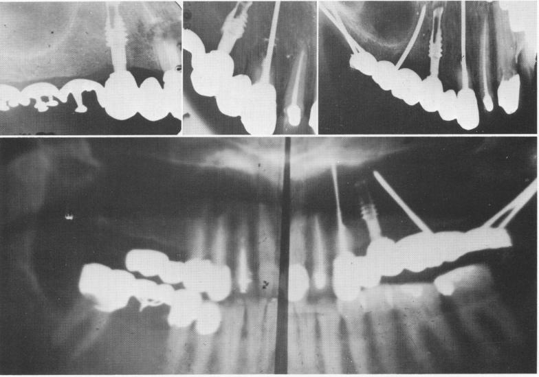
Fig. 13-56. A, Roentgenogram shows broken cuspid root prior to its removal. The vent-plant is seen in the bicuspid region. B, The endodontic implant stabilizer as seen 6 months after apicoectomy. C, A cross-sectional occlusal x-ray taken 2 years after initial insertions shows the endodontic implant stabilizer, the vent-plant, and the triplant. Notice the regeneration of bone resembling a lamina dura around the mesial wall of the vent plant and the circumvention of the sinus with the triplant. D, A Panorex x-ray illustrating the bridge and implants 5 years postoperatively.
B
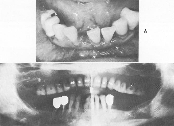
Fig. 13-57. A, The existing teeth were extremely loose. B, A Panorex shows very little sup-porting bone.
|
|
Page 603 |
Next Page |
|
Copyright warning: This information is presented here for free for anyone to study online. We own exclusive internet copyrights on all content presented on this website. We use sophisticated technology to identify and legally close down websites that reproduce copyrighted content without permission - so please don’t do it.
|