| Theories and Techniques of Oral Implantology (vol.2) (published 1970) | Dr. Leonard I. Linkow |
|
|
Next Page |
| This is an archival HTML version of this book originally hosted here in 2006. The HTML may not display well on modern browsers. Please view the modern PDF Version for a better viewing experience. |
602 Theories and techniques of oral implantology
A
B
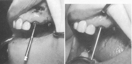
Fig. 13-52, A, The gold post was removed from the fractured cuspid, and the apical half of the root was removed through the labial cortical plate. Rotary reamers were then used to widen the intact portion of the root. B, The endodontic root stabilizer was then fitted accurately inside the root, extending deep beyond the void left by the removed root.
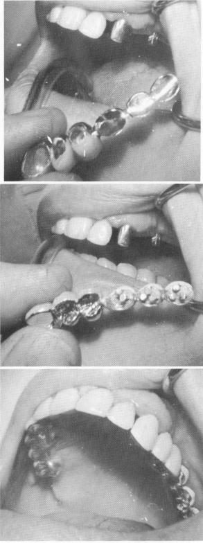
Fig. 13-53. A, The bridge with the scalloped template was fitted into position. B and C, The prosthesis was cemented with hard cement.
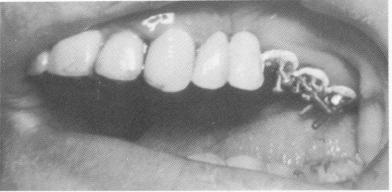
Fig. 13-54. Pin implants were driven through the openings in the template.
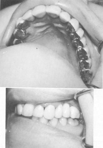
Fig. 13-55. The superstructure was cemented over the template after the triplant pins were affixed with acrylic.
A
B
C
|
|
Page 602 |
Next Page |
|
Copyright warning: This information is presented here for free for anyone to study online. We own exclusive internet copyrights on all content presented on this website. We use sophisticated technology to identify and legally close down websites that reproduce copyrighted content without permission - so please don’t do it.
|