| Theories and Techniques of Oral Implantology (vol.2) (published 1970) | Dr. Leonard I. Linkow |
|
|
Next Page |
| This is an archival HTML version of this book originally hosted here in 2006. The HTML may not display well on modern browsers. Please view the modern PDF Version for a better viewing experience. |
Endodontic implants 595
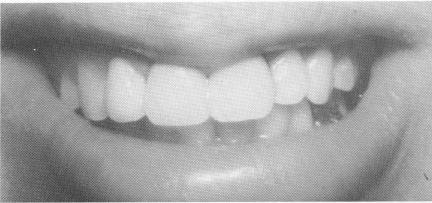
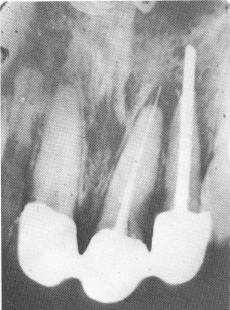
Fig. 13-25. The three-unit completed porcelain-fused-tometal splint helped to support the reimplanted tooth.
horizontally below the tongue. Eighteen hours after the accident, the luxated teeth were realigned and the knocked-out ones reimplanted; all of them were stabilized with endodontic implants. No other splints were used. The patient was pain-free and could eat normally 1 hour after the operation. Some years later resorption started on the reimplanted teeth. Eventually the roots resorbed, leaving only the stabilizers. These were still firmly fixed in the bone without any roots around them. This apparently indicates that the diseased root acts as an irritant and is eliminated by the body, but the implant is tolerated even as nearby diseased tissues are re-sorbed.
So far it has not been possible to predict or to exclude the onset of root resorption, nor has it been possible to foretell the type of resorption. Two types of root resorption have been observed. One type commences at the apex and slowly destroys the root. This process is quite painless. The other type of resorption seems to be initiated by an aggressive metaplasia of the marginal gums, which eventually severs the tooth into two parts. Here the gums are painful and bleeding.
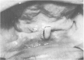
Fig. 13-27. The only teeth remaining were a loose lateral incisor and second molar.
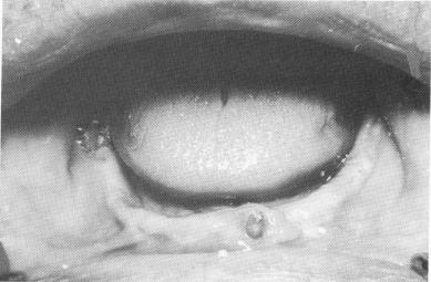
Fig. 13-28. The lateral incisor and molar were prepared for full coverage.
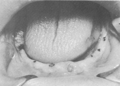
Fig. 13-29. All the contemplated implant sites were marked with indelible pencil.
ENDODONTIC ROOT STABILIZERS COMBINED WITH PROSTHODONTIC IMPLANTS
In combination with endosseous and subperiosteal implants, endodontic pin implants are used primarily to stabilize loose teeth to serve as natural abutments for fixed dentures. As always, the
Fig. 13-26. Postoperative radiograph.
|
|
Page 595 |
Next Page |
|
Copyright warning: This information is presented here for free for anyone to study online. We own exclusive internet copyrights on all content presented on this website. We use sophisticated technology to identify and legally close down websites that reproduce copyrighted content without permission - so please don’t do it.
|