| Theories and Techniques of Oral Implantology (vol.2) (published 1970) | Dr. Leonard I. Linkow |
|
|
Next Page |
| This is an archival HTML version of this book originally hosted here in 2006. The HTML may not display well on modern browsers. Please view the modern PDF Version for a better viewing experience. |
596 Theories and techniques of oral implantology
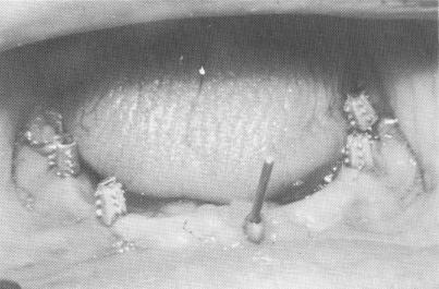
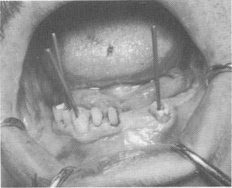
Fig. 13-30. An endodontic root stabilizer was set deeply into the bone existing below the loose incisor, immediately stabilizing it. All implants were set into the bone, and gold copings were placed over them.
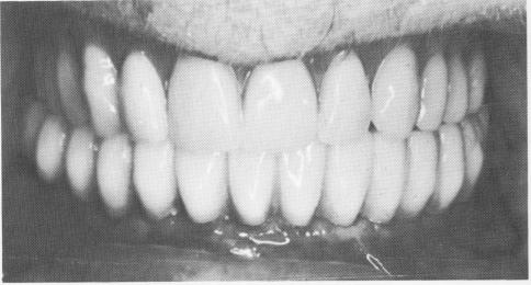
Fig. 13-31. A full arch fixed denture was cemented over the abutments.
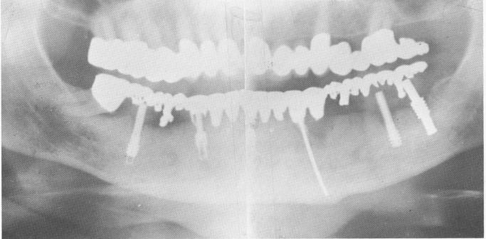
Fig. 13-32. A 3-year postoperative Panorex roentgenogram shows the endodontic implant stabilizer and the implants. Some bone resorption is evident around the left first implant extending downward from the alveolar crest. Also, an infrabony pocket exists along part of the mesial wall of the remaining right molar. The patient, however, has been enjoying eating and chewing for the past 3'/rz years. There is no mobility.
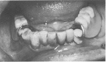
Fig. 13-33. The mouth of a 55-year-old physician.
Fig. 13-34. Three loose teeth were supported with endodontic stabilizers.
|
|
Page 596 |
Next Page |
|
Copyright warning: This information is presented here for free for anyone to study online. We own exclusive internet copyrights on all content presented on this website. We use sophisticated technology to identify and legally close down websites that reproduce copyrighted content without permission - so please don’t do it.
|