| Theories and Techniques of Oral Implantology (vol.2) (published 1970) | Dr. Leonard I. Linkow |
|
|
Next Page |
| This is an archival HTML version of this book originally hosted here in 2006. The HTML may not display well on modern browsers. Please view the modern PDF Version for a better viewing experience. |
Subperiosteal implants 559
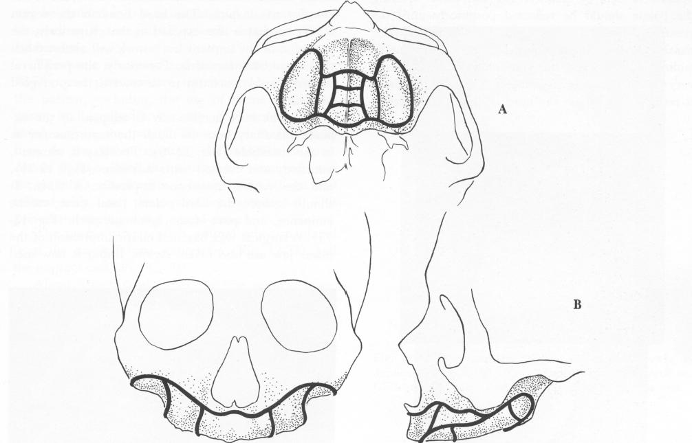
Fig. 12-70. A, The design of the palatal portion of the subperiosteal maxillary implant must be as broad as possible (Linkow). B, The peripheral borders should end close to the base of the anterior nasal spine, extend high into the canine eminence and slightly below the infraorbital canals, and extend further distally along the zygomatic arch and along the lateral wall of the pterygoid plate.
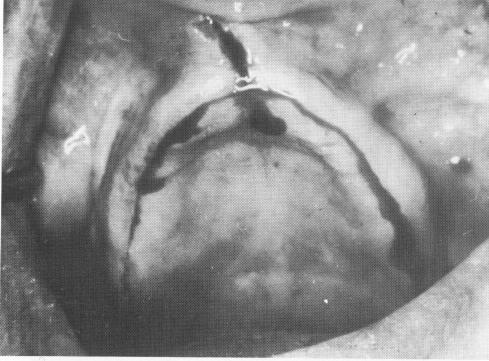
Fig. 12-71. An incision is made along the crest of the ridge from maxillary tuberosity to maxillary tuberosity. Some-times a vertical accessory incision is made; this can be seen in the anterior region above the primary incision.
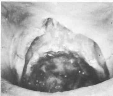
Fig. 12-72. The fibromucosal tissue on the labial and buccal aspect is retracted to expose the base of the anterior nasal spine as well as the canine eminence, zygomatic arch, and pterygoid process. The palatal tissue is retracted to expose the entire hard palate, and sometimes the posterior nasal spine and greater and lesser palatine foramina.
|
|
Page 559 |
Next Page |
|
Copyright warning: This information is presented here for free for anyone to study online. We own exclusive internet copyrights on all content presented on this website. We use sophisticated technology to identify and legally close down websites that reproduce copyrighted content without permission - so please don’t do it.
|