| Theories and Techniques of Oral Implantology (vol.2) (published 1970) | Dr. Leonard I. Linkow |
|
|
Next Page |
| This is an archival HTML version of this book originally hosted here in 2006. The HTML may not display well on modern browsers. Please view the modern PDF Version for a better viewing experience. |
Endosseous blade implants 511
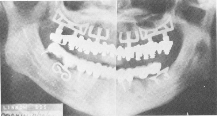
Fig. 11-158. The final Panorex. An apicoectomy had been done at the apex of the lower left first bicuspid tooth, and a tooth extraction was done 1 week before the radiograph was taken. (From Linkow, L. I.: Status of oral implants, 1969, Inform. Odontostomat., Vol. 1, 1969.)
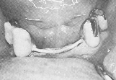
Fig. 11-159. Preoperative view of patient's existing teeth and prosthesis.
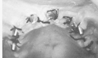
Fig. 11-160. Blade implants were placed in the edentulous maxilla and sutured.
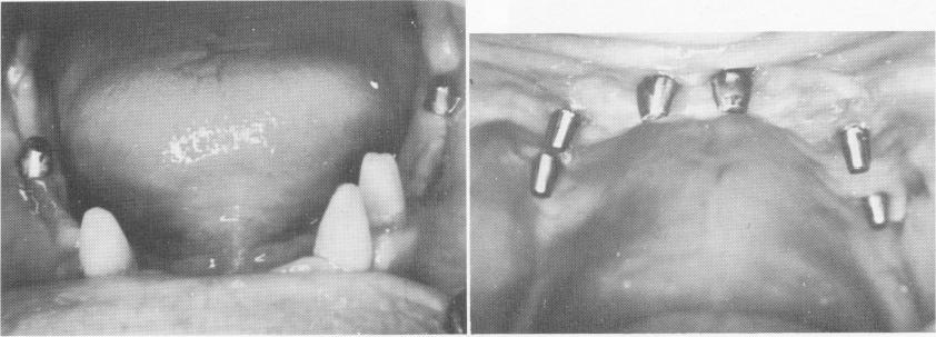
Fig. 11-161. When the tissues had healed, impressions were taken for the prostheses.
|
|
Page 511 |
Next Page |
|
Copyright warning: This information is presented here for free for anyone to study online. We own exclusive internet copyrights on all content presented on this website. We use sophisticated technology to identify and legally close down websites that reproduce copyrighted content without permission - so please don’t do it.
|