| Theories and Techniques of Oral Implantology (vol.2) (published 1970) | Dr. Leonard I. Linkow |
|
|
Next Page |
| This is an archival HTML version of this book originally hosted here in 2006. The HTML may not display well on modern browsers. Please view the modern PDF Version for a better viewing experience. |
510 Theories and techniques of oral implantology
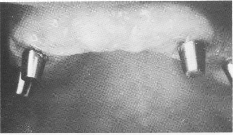
Fig. 11-155. A close-up view of the healed tissues before the final prosthesis was inserted.
the implant posts. It was then carefully occluded with the lower jaw.
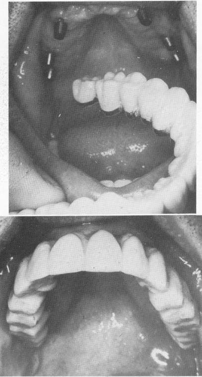
When complete healing had taken place (Fig. 11-153), interchangeable prefabricated gold copings were placed over the implant posts. A bite registration and a full mouth plaster index were taken, picking up the copings.
At the next visit, the soldered full arch splint was tried in the mouth (Fig. 11-154). A final wax inter-occlusal record of centric relation was then taken for completing the bridge.
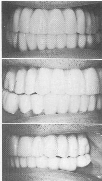
By the time the bridge was ready for final insertion, the tissues around the posts were completely healed and closely adapted (Fig. 11-155). The full arch fixed denture, made of acrylic-over-gold, was cemented over the metal posts with hard cement (Fig. 11-156). The occlusion was then carefully spot-ground for any other prematurities caused by the addition of the cement (Fig. 11-157).
Fig. 11-156. The final prosthesis in place.
Fig. 11-157. The final prosthesis was articulated.
|
|
Page 510 |
Next Page |
|
Copyright warning: This information is presented here for free for anyone to study online. We own exclusive internet copyrights on all content presented on this website. We use sophisticated technology to identify and legally close down websites that reproduce copyrighted content without permission - so please don’t do it.
|