| Theories and Techniques of Oral Implantology (vol.1) (published 1970) | Dr. Leonard I. Linkow |
|
|
Next Page |
| This is an archival HTML version of this book originally hosted here in 2006. The HTML may not display well on modern browsers. Please view the modern PDF Version for a better viewing experience. |
Implant histology 103
severed from one another. The implant in the nasal septum was loose and easily removed (Fig. 4-40). The other vent-plants were extremely firm and were removed so as to include bone blocks (Fig. 4-41) .
Histologic reports from Dr. George Greene, Jr., of the University of Buffalo, Buffalo, New York, revealed that the tissue adjacent to the bone-encapsulated implants was fibrous in nature, with the fiber content predominating and with a small amount of
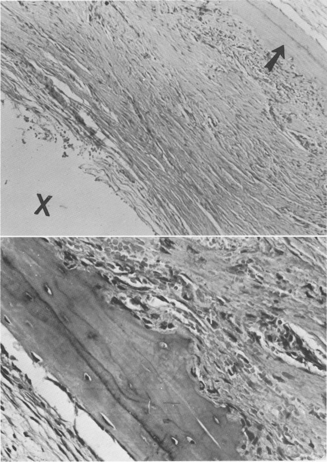
cellularity (Fig. 4-42). Deeper in the tissue around each implant was a ring of bone. The side toward the implants exhibited active osteogenesis, indicating deposition of bone toward the implant site (Fig. 4-43). Other studies at this deeper level showed the inner aspect of the fibrous tissue as it butted against the implants (Fig. 4-44) . On the whole, fiber elements predominated over the cellular elements. However, near the implant there were more cells and the fiber
Fig. 4-43. A, In addition to the collagenous pseudoperiodontal membrane around the implant site (X), osteogenesis of the bone is seen (arrow) with the osteoblasts lined up toward the implant site. B, Under higher power, osteocytes are clearly visible in their lacunae.
|
|
Page 103 |
Next Page |
|
Copyright warning: This information is presented here for free for anyone to study online. We own exclusive internet copyrights on all content presented on this website. We use sophisticated technology to identify and legally close down websites that reproduce copyrighted content without permission - so please don’t do it.
|