| Theories and Techniques of Oral Implantology (vol.1) (published 1970) | Dr. Leonard I. Linkow |
|
|
Next Page |
| This is an archival HTML version of this book originally hosted here in 2006. The HTML may not display well on modern browsers. Please view the modern PDF Version for a better viewing experience. |
104 Theories and techniques of oral implantology
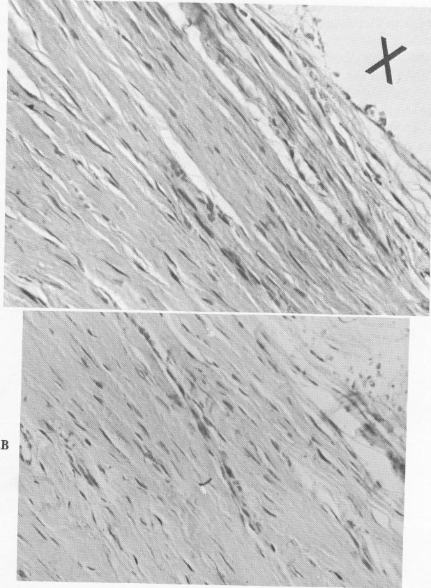
Fig. 4-44. A, Under 200x magnification, fibrous elements and cells are visible. There is a higher percentage of fibroblastic cells nearer the implant site (X). B, The cells and fibers are regularly arranged, characteristic of a healthy site.
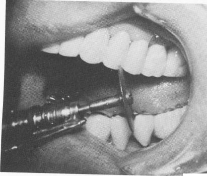
Fig. 4-45. To remove the vent-plants, it was necessary to disk away part of the splint.
|
|
Page 104 |
Next Page |
|
Copyright warning: This information is presented here for free for anyone to study online. We own exclusive internet copyrights on all content presented on this website. We use sophisticated technology to identify and legally close down websites that reproduce copyrighted content without permission - so please don’t do it.
|