| Theories and Techniques of Oral Implantology (vol.1) (published 1970) | Dr. Leonard I. Linkow |
|
|
Next Page |
| This is an archival HTML version of this book originally hosted here in 2006. The HTML may not display well on modern browsers. Please view the modern PDF Version for a better viewing experience. |
Implant histology 105
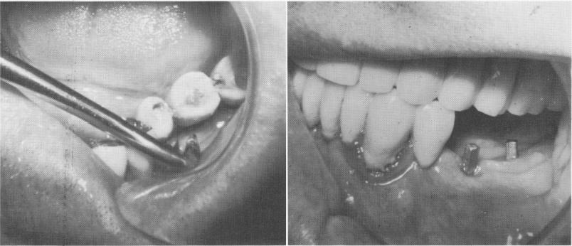
Fig. 4-46. The implants.
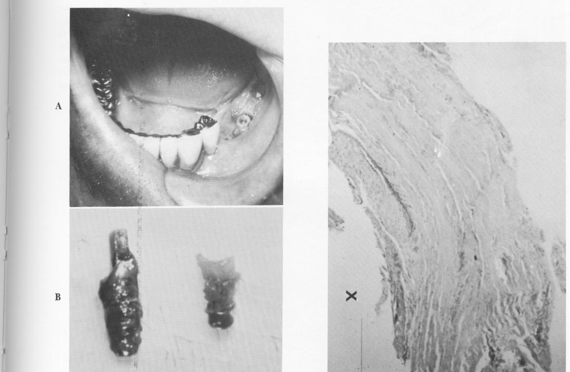
r
Fig. 4-47. The anterior implant was easily unscrewed. The posterior implants were removed by drilling out a circular core of bone surrounding each one. B, The removed implants. That on the right was so firmly embedded that it broke during removal.
Fig. 4-48. A cross-sectional low power magnification shows that the soft tissue follows the contour of the implant site (X).
|
|
Page 105 |
Next Page |
|
Copyright warning: This information is presented here for free for anyone to study online. We own exclusive internet copyrights on all content presented on this website. We use sophisticated technology to identify and legally close down websites that reproduce copyrighted content without permission - so please don’t do it.
|