| Theories and Techniques of Oral Implantology (vol.1) (published 1970) | Dr. Leonard I. Linkow |
|
|
Next Page |
| This is an archival HTML version of this book originally hosted here in 2006. The HTML may not display well on modern browsers. Please view the modern PDF Version for a better viewing experience. |
106 Theories and techniques of oral implantology
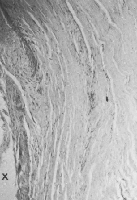
Fig. 4-49. Fibroblasts are evident under higher magnification.
content was less marked than further away from the implant. This is consistent with the variations in cellularity observed in the periodontal membrane.
Case 2. Another opportunity for careful histologic studies arose when a patient's implants had to be removed because of paresthesia. The 49-yearold male patient wore a full lower fixed denture supported by two cuspid teeth and six endosseous implants. A paresthesia developed on the lower left side because the implants in that area had been set too deeply. The symptoms continued after onset for 14 months, until the last three implants on the lower left side were removed.
To get at the implants, the posterior quadrant on the left side of the splint was disked away from the anterior portion (Fig. 4-45). It was then removed from the underlying implants (Fig. 4-46). The first implant was removed by simply unscrewing it, but the second implant was so firmly fixed that it broke at the alveolar crest. A complete bony circle had to be drilled around it to remove it (Fig. 4-47). The most posterior implant was also removed with bone encircling it. Only the implants with bone were sent to the laboratory. The histologic report by George Greene, Jr., of the University of Buffalo, follows:
GROSS: Two threaded metal implant screws were received and one small, separate calcified fragment. The largest screw measured 17 x 5 x 4 mm., and contained a moderate amount of tena-
2 s-SOW. 119,11,
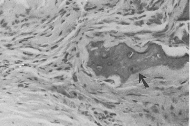
Fig. 4-50. Small area of calcification (arrow) near the periphery of the soft tissue strip.
|
|
Page 106 |
Next Page |
|
Copyright warning: This information is presented here for free for anyone to study online. We own exclusive internet copyrights on all content presented on this website. We use sophisticated technology to identify and legally close down websites that reproduce copyrighted content without permission - so please don’t do it.
|