| Theories and Techniques of Oral Implantology (vol.1) (published 1970) | Dr. Leonard I. Linkow |
|
|
Next Page |
| This is an archival HTML version of this book originally hosted here in 2006. The HTML may not display well on modern browsers. Please view the modern PDF Version for a better viewing experience. |
Implant histology 107
cious fibrous tissue obstructing the screw threads. The second screw was shorter, measuring 7 x 5 x 4 mm. and was missing the post portion. The screw portion had a small amount of tissue obstructing the threads. The separate calcified fragment was ovoid in shape and measured 4 x 4 x 3 mm.
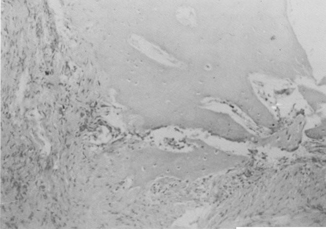
MICROSCOPIC DESCRIPTION: The specimens con-
sist of strips of connective tissue. In some areas the strips assume a semilunar configuration rep-resenting tissue which was in direct contact with the implant screws [Fig. 4-48]. This tissue is comprised of well-developed mature collagen bundles and is devoid of inflammatory cells. Foci of proliferating plump fibroblasts are evident [Fig. 4-49;, and in one area small calcifications are
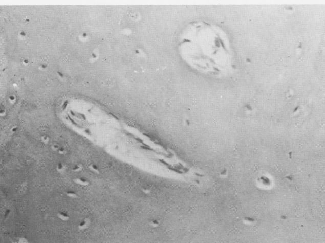
Fig. 4-51. That part of the soft tissue specimen away from the implant site contains numerous islands of bone.
Fig. 4-52. Under higher magnification the bone fragments are seen to contain numerous osteocytes.
|
|
Page 107 |
Next Page |
|
Copyright warning: This information is presented here for free for anyone to study online. We own exclusive internet copyrights on all content presented on this website. We use sophisticated technology to identify and legally close down websites that reproduce copyrighted content without permission - so please don’t do it.
|