| Theories and Techniques of Oral Implantology (vol.1) (published 1970) | Dr. Leonard I. Linkow |
|
|
Next Page |
| This is an archival HTML version of this book originally hosted here in 2006. The HTML may not display well on modern browsers. Please view the modern PDF Version for a better viewing experience. |
120 Theories and techniques of oral implantology
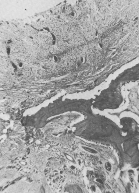
The results when the opening was placed too high coronally are different (Fig. 4-78). The osseous bridge that began to form underwent an inflammatory breakdown analogous to a periodontal abscess. Under higher power magnification, the inflammatory reaction is clearly seen (Fig. 4-79). From these results, one may conclude that osseous bridges grow through openings in the roots of the tooth replica polymer implant. These openings must not be placed too high coronally but should be placed in the lower third of the implant's root if the osseous bridge is to remain healthy.
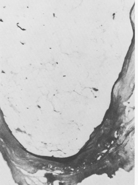
The fate of the tissues directly adjacent to the implant material was very promising in some cases. There was good periodontal tissue adaptation composed of regular, well-formed connective tissue.
Fig. 4-80 demonstrates that the connective tissues of the "periodontal" membrane extended into the implant material. Under progressively higher snag-
% ^'+ i.pOt ►t,
nification these clearly appear to be extensions of the periodontal ligament (Fig. 4-81) .
In summary, Hodosh and his associates found osteoblastic activity in the alveolar bone directly adjacent to the pseudoperiodontal membrane around the plastic implant. The fibers of this membrane tended to run in a more vertical direction than with a normal tooth. However, at times these fibers were seen to insert directly into the plastic material. In implants with holes near their apices, the implants were extremely solid within their sockets after 6 to 12 months. They could he removed only by vertical incisions and sectioning of one end of the horizontal bony bridge. Varying, but minimal, degrees of inflammation of the gingival tissue occurred around the plastic implants.
Buhite on Linkow's blade implant
The first histologic report on a blade implant came from Robert Buhite, D.D.S., of Rochester, New
Fig. 4-79. The inflammation and breakdown are more clearly seen under higher magnification. (Courtesy M. Hodosh.)
Fig. 4-80. Even under low power extensions of the connective tissue into the implant are evident. (Courtesy M. I Iodosh. )
|
|
Page 120 |
Next Page |
|
Copyright warning: This information is presented here for free for anyone to study online. We own exclusive internet copyrights on all content presented on this website. We use sophisticated technology to identify and legally close down websites that reproduce copyrighted content without permission - so please don’t do it.
|