| Theories and Techniques of Oral Implantology (vol.1) (published 1970) | Dr. Leonard I. Linkow |
|
|
Next Page |
| This is an archival HTML version of this book originally hosted here in 2006. The HTML may not display well on modern browsers. Please view the modern PDF Version for a better viewing experience. |
Implant histology 121
York. The patient, a German woman in her late fifties, had for 9 months enjoyed a successful blade implantation. However, she was unexpectedly returning to her homeland permanently and feared that if any problems arose, there would be no one to take care of them. For this reason, she requested removal of the implant.
When the bridge was removed to get at the implant, the tissues under it appeared pink, firm, and healthy (Fig. 4-82). To remove the implant itself, Dr. Buhite made three vertical incisions along the entire anteroposterior length of the blade: one along the alveolar crest through the soft tissues down to the shoulders of the blade, one on the buccal face of
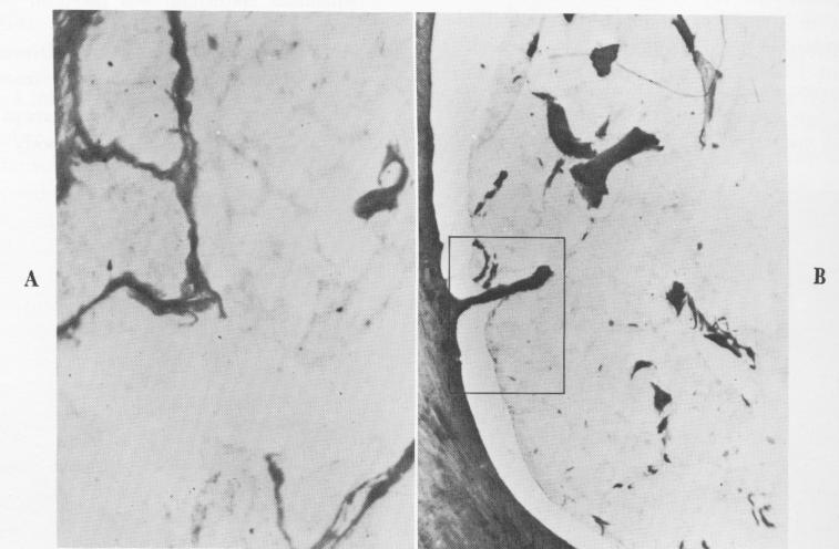
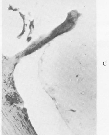
Fig. 4-81. A, High-power magnification of the interconnected strands extending from the fibrous membrane into the implant. B, A remarkably clear demonstration of a fibrous extension into the implant. C, Under even higher power, the nature of the extension is demonstrated. (Courtesy M. Hodosh.)
|
|
Page 121 |
Next Page |
|
Copyright warning: This information is presented here for free for anyone to study online. We own exclusive internet copyrights on all content presented on this website. We use sophisticated technology to identify and legally close down websites that reproduce copyrighted content without permission - so please don’t do it.
|