| Theories and Techniques of Oral Implantology (vol.1) (published 1970) | Dr. Leonard I. Linkow |
|
|
Next Page |
| This is an archival HTML version of this book originally hosted here in 2006. The HTML may not display well on modern browsers. Please view the modern PDF Version for a better viewing experience. |
122 Theories and techniques of oral implantology
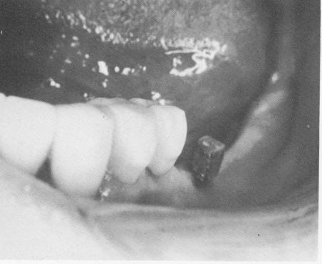
the blade down to its inferior border, and one on the lingual face down to the implant's inferior border (Fig. 4-83).
The bone completely covered the superior surface of both shoulders and had to be removed with a round bur.
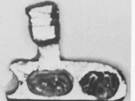
When the blade was removed, there were two plugs of bone firmly affixed inside the vents (Fig. 4-84). The pathologic report of Edmund Cataldo, D.D.S., M.S., of the Department of Oral Pathology, Tufts University, Boston, follows:
MICROSCOPIC DESCRIPTION: Decalcification sec-
tions show two irregular and fragmented masses of bone with marrow and fibrous connective tissue.
The bone appears normal in configuration and evidence of osteoblastic activity is noted. A granular black foreign material evoking a foreign body giant cell reaction is noted in the connective tissue and marrow spaces. Also noted in the connective tissue is a mild to moderate chronic inflammatory infiltrate consisting of plasma cells, lymphocytes, and histiocytes. In examining all tissue sections, a small fragment of stratified squamous epithelium was noted in proximity to bone.
MICROSCOPIC, DIAGNOSIS: Bone reveals chronic
inflammation and a foreign body reaction.
The report was satisfactory except for the evidence of inflammatory foreign body reaction and
Fig. 4-82. The soft tissues around a blade implant successfully functioning for 9 months appear to be firm and healthy. (Courtesy R. Buhite.)
Fig. 4-83. Three incisions were necessary to free the firmly affixed blade. (Courtesy R. Buhite.)
Fig. 4-84. The blade with bone in its vents. (Courtesy R. Buhite.)
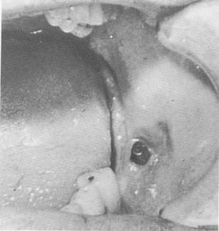
Fig. 4-85. The protruding post of the blade was irritating the patient's cheek because it was originally inserted too far buccally.
|
|
Page 122 |
Next Page |
|
Copyright warning: This information is presented here for free for anyone to study online. We own exclusive internet copyrights on all content presented on this website. We use sophisticated technology to identify and legally close down websites that reproduce copyrighted content without permission - so please don’t do it.
|