| Theories and Techniques of Oral Implantology (vol.1) (published 1970) | Dr. Leonard I. Linkow |
|
|
Next Page |
| This is an archival HTML version of this book originally hosted here in 2006. The HTML may not display well on modern browsers. Please view the modern PDF Version for a better viewing experience. |
146 Theories and techniques of oral implantology
soft tissues. By the time bone regenerates between the posts from the apical portion of the implant, a great deal of soft tissue will have invaginated from the oral mucosa and filled inside the uprights. Thus bone cannot fill in and stabilize the implant, and it will fail. In addition, the smooth sides provide no surface variation for connective tissue involvement.
Pretto tried another approach quite different from his trombone design. Unlike the single-unit trombone, the adjustable winglet implant is composed of three parts (Fig. 5-17) . The principal part is a post, the bottom of which is flared and contains openings through which bone can grow. The upper-most part of the post is threaded. A tantalum band that forms winglets is screwed over the threaded section. These extremely thin winglets contain windows through which bone can grow. Over the band is screwed a disk that causes the winglets to flare until they touch the sides of the cavity created for the implant. It is hoped that bracing the implant will hold it secure until bone regrows around and through the other parts of the implant.
The idea, although mechanically practical, does not take into account that pressure causes bone resorption. Unless the operator flares the winglets until they barely touch the walls of the socket, he is creating a problem that will enlarge the resorption zone and cause eventual loosening of the implant.
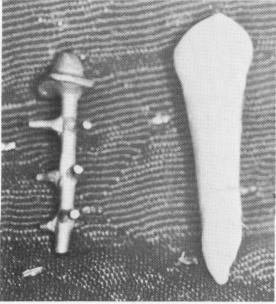
One of the more successful post designs is that of the Chinese-American Ted Lee (Fig. 5-18) . The central post is narrow, with small extensions that pro-vide security against exfoliation. The spacing between the extensions and the narrowness of the post allow blood- and bone-building elements to encompass the major part of the implant.
Fig. 5-18. The protruding extensions of this Lee implant give the implant the same diameter as the incisor which it is to replace. (Courtesy T. C. Lee.)
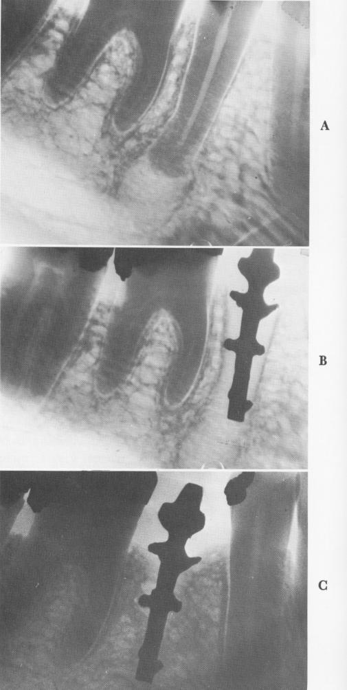
Fig. 5-19. A, A large granuloma is found around the apex of the second bicuspid to be replaced by a Lee implant. B, Some bone regrowth is seen 9 months after the implant's insertion into the open socket. C, Thirteen years later, the implant is still quite secure and functioning well, with bone practically totally surrounding the implant. (Courtesy T. C. Lee.)
|
|
Page 146 |
Next Page |
|
Copyright warning: This information is presented here for free for anyone to study online. We own exclusive internet copyrights on all content presented on this website. We use sophisticated technology to identify and legally close down websites that reproduce copyrighted content without permission - so please don’t do it.
|