| Theories and Techniques of Oral Implantology (vol.1) (published 1970) | Dr. Leonard I. Linkow |
|
|
Next Page |
| This is an archival HTML version of this book originally hosted here in 2006. The HTML may not display well on modern browsers. Please view the modern PDF Version for a better viewing experience. |
314 Theories and techniques of oral implantology
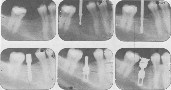
Fig. 8-8. Intraoral radiographs illustrating the step-by-step technique. (From Linkow, L. I.: The age of endosseous implants, Dent. Concepts, Spring, 1966.)
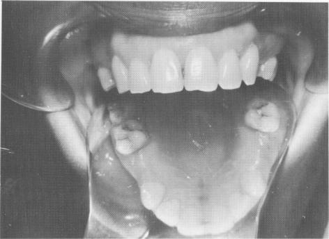
Fig. 8-9. Two bicuspids were missing bilaterally in an otherwise caries-free maxilla. Also, no previous fillings existed.
sure that the occlusion is correct from the moment of insertion.
As is important in all implant interventions, radiographs were taken before, during, and after implant insertion (Fig. 8-8).
Case 2
Ligated single tooth implants
Here single tooth restorations were performed on a 32-year-old woman who was missing both maxillary bicuspid teeth on both sides of her arch (Fig. 8-9). Because her neighboring teeth had no fillings or decay, she did not want them prepared as abutments.
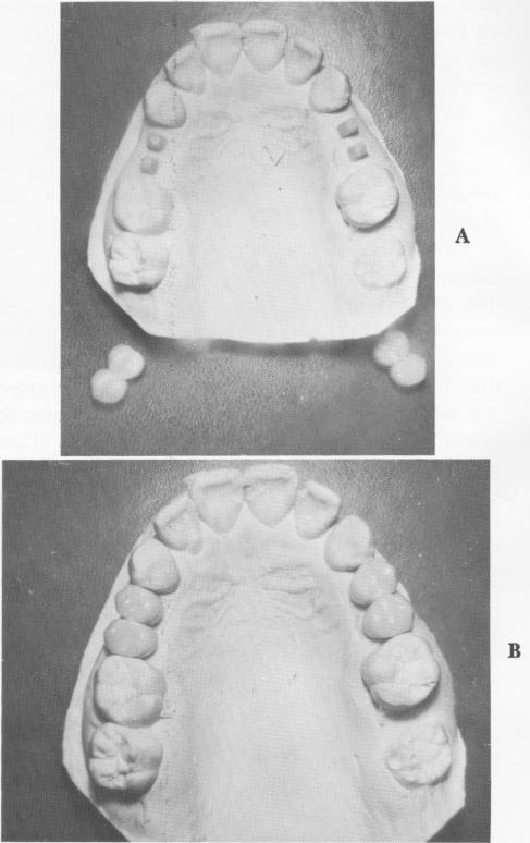
An alginate impression of her entire maxillary arch was taken, as well as a wax interocclusal record
Fig. 8-10. A, Two prefabricated acrylic-over-gold splints were made for both edentulous areas using duplicate implant shafts coated with nailpolish to build them up. B, The restorations fitted over the duplicate shafts.
|
|
Page 314 |
Next Page |
|
Copyright warning: This information is presented here for free for anyone to study online. We own exclusive internet copyrights on all content presented on this website. We use sophisticated technology to identify and legally close down websites that reproduce copyrighted content without permission - so please don’t do it.
|