| Theories and Techniques of Oral Implantology (vol.1) (published 1970) | Dr. Leonard I. Linkow |
|
|
Next Page |
| This is an archival HTML version of this book originally hosted here in 2006. The HTML may not display well on modern browsers. Please view the modern PDF Version for a better viewing experience. |
Implant histology 93
Linkow on Chercheve's spiral-post implant
As was stated earlier in this chapter, the opportunity of obtaining for study human bone specimens containing implants is rare. Linkow was quite fortunate in this respect and was able to get some bone blocks from a few patients. In two cases given here, the implants had been functioning successfully for a period of 3 years and 212 years, respectively. The situations providing bone blocks containing successful implants are worth examining in detail.
Case 1. The first case involves a man healthy in all respects. In May, 1962, two of his lower left bicuspids were prepared as the anterior abutments for a fixed partial denture that would utilize two molar endosseous implants as the posterior abutments. At the second visit, the two implants were set into the bone; a prefabricated temporary acrylic splint was placed over the two prepared tooth abutments anteriorly and over the two implant abutments posteriorly. The next day the patient came into the office very much aggravated, demanding that his "painful" implants be removed. When the situation was examined more carefully, it appeared that it was not the implants that were bothering him but rather his second bicuspid, which had been pre-pared the previous week and had flared up. After much convincing, the patient finally allowed the removal of the nerve tissue of the second bicuspid. The patient was told to report back the next day, regardless of his symptoms. If any pain persisted, the implants would be removed.
Two weeks later the patient reappeared for the first time, claiming that he had been pain-free and that the implant prosthesis felt just like his own teeth. He enthusiastically requested a permanent implant bridge for his upper teeth also, as well as a mandibular unilateral fixed partial denture for his right side. He only had his maxillary anterior eight teeth present, from first bicuspid to first bicuspid. The maxillary sinus floors on both sides were very high, providing an unusual amount of bone between the sinus floors and the alveolar ridges. Five Chercheve spiraled implants were set in the posterior edentulous areas on both sides of the maxilla, and a full arch fixed denture of acrylic and gold was fabricated. Roentgenograms showing these, as well as the mandibular implants, in place are shown in Fig. 4-18.
The implants remained in the mouth until July, 1965, 3 years later. After this long lapse in time the patient, who had been carefully examined about every 3 months from the time of insertion, began to insist that the implants, although functioning well,
be removed. Finally, after much persuasion to leave them in, he confessed that he had been told that he could get into a lot of "serious trouble" with implants and could even get cancer. He was frightened and adamant that they be removed.
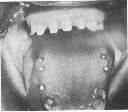
Roentgenograms and examination illustrated that all of the implants were firmly fixed deeply into the osseous tissue and that there were no pathologic areas (Fig. 4-19). There was no pain on palpation. Although the patient was completely comfortable, his implants were to be removed. Here was an ideal case for bone sections. After carefully removing the bridges, the tissues around the implants were found to be firm, healthy, and closely adapted to the implants (Fig. 4-20). The implants were removed by disking and cutting away part of the surrounding alveolar bone (Fig. 4-21) .
The following is a report from Harry Lane Robinson, M.D., Professor of Pathology, New York University School of Dentistry, on the histologic sections (Fig. 4-22) :
Examination of the portions of bone submitted with screws in situ reveals that the bone is viable and the marrow spaces are filled with normal adipose marrow. There is no evidence of necrosis or inflammation. Several sections at various levels through the specimens were examined. It would seem that the screws are well tolerated by the bone and apparently do not cause reactions.
There is a small portion of mucous membrane attached to a bony spicule which is edematous, congested, and contains a moderate number of lymphocytes and monocytes.
Fig. 4-20. Clinical view with the bridge removed. Note the healthy tissues under the pontics around the implant posts.
|
|
Page 93 |
Next Page |
|
Copyright warning: This information is presented here for free for anyone to study online. We own exclusive internet copyrights on all content presented on this website. We use sophisticated technology to identify and legally close down websites that reproduce copyrighted content without permission - so please don’t do it.
|