| Theories and Techniques of Oral Implantology (vol.1) (published 1970) | Dr. Leonard I. Linkow |
|
|
Next Page |
| This is an archival HTML version of this book originally hosted here in 2006. The HTML may not display well on modern browsers. Please view the modern PDF Version for a better viewing experience. |
94 Theories and techniques of oral implantology
B
C
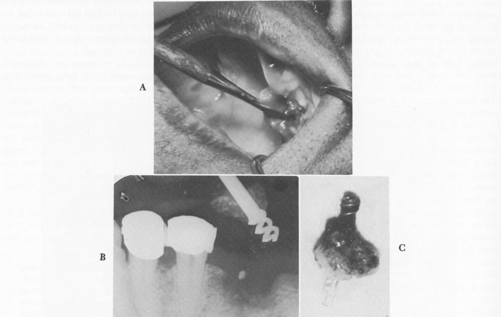
Fig. 4-21. A, The implants were removed by cutting away alveolar bone. B, Radiograph of a lower implant during removal. C, The specimen, a successfully implanted Chercheve spiral-post implant surrounded by bone.
Case 2. In the second case, the reason for removing three endosseous implants from the maxilla of a healthy, 50-year-old woman was unique. The patient had no teeth in the right maxilla from the central incisor to the tuberosity; most of her teeth were present on the left side (Fig. 4-23). The patient wore a removable partial denture and was desperate to have a fixed full arch denture fabricated for esthetic reasons and function. There existed, however, a completely horizontal maxillary cuspid impaction on her right side that was intact with the floor of the antrum (Fig. 4-24).
Removing the impacted tooth presented a problem. There was practically no alveolar bone between the tooth and the ridge. If the tooth were removed, the little bone remaining would be lost and complete distortion would occur on that side. Another indication that the tooth should remain undisturbed was that the patient had always been pain-free in that area.
In February, 1963, the implantation procedure was begun. The teeth on the left side were prepared for full crown preparations, impressions were taken of them, and castings were fabricated and fitted over the teeth. Three spiral type implants were set into the edentulous right side of the maxilla. The maxillary tuberosity area distal to the impaction was utilized for the placement of two implants, and the third was placed under the crown of the cuspid (Fig. 4-25). A final wax bite and plaster index were taken for the fabrication of a full arch, acrylic-andgold, fixed denture, which was cemented over the teeth and implants with oxyphosphate of zinc cement (Fig. 4-26). The occlusion was carefully checked and balanced (Fig. 4-27). The patient was told that if the cuspid ever "acted up" the implants would have to be removed.
Two years later, in 1965, the cuspid impaction became troublesome and required removal. In order to get at the cuspid, a portion of the bridge had to
|
|
Page 94 |
Next Page |
|
Copyright warning: This information is presented here for free for anyone to study online. We own exclusive internet copyrights on all content presented on this website. We use sophisticated technology to identify and legally close down websites that reproduce copyrighted content without permission - so please don’t do it.
|