| Theories and Techniques of Oral Implantology (vol.1) (published 1970) | Dr. Leonard I. Linkow |
|
|
Next Page |
| This is an archival HTML version of this book originally hosted here in 2006. The HTML may not display well on modern browsers. Please view the modern PDF Version for a better viewing experience. |
Implant histology 99
bone cuttings, possibly remnants of curetting or of a bone bur. These are not remarkable. A few small chronic inflammatory cells are encountered in one area, but there is no significant infiltration. There can be seen dense active bone with an irregular growth pattern. There is no evidence of malignancy.
As an addendum, the pathologist drew some generalizations.
In reviewing the cases submitted by Dr. Linkow to the laboratory through your office, we made several observations: Production of fibrous tissue around the metal posts appears to be a fairly uniform reaction. Much of this fibrous tissue is probably functional, although it does allow more movement than intact and well-adapted bone. Inflammatory involvement of the fibrous tissue is primarily associated with proximity to the crevicular areas.
The extension of crevicular epithelium and the presence of other chronic inflammation adja-
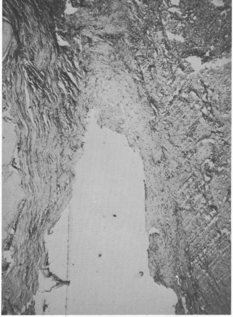
Fig. 4-32. A section from a failing implant. Note the in-filtration of inflammatory cells and the extra thickened granulation tissue.
cent to this epithelium was a rather constant feature. It has been my experience that the gingival cuff around a metal post is invariably a problem from this standpoint.
The tenacity of the fibrous tissue away from the crevicular area was rather remarkable. This feature has also been observed around wires and pins placed within the bone adjacent to fractures. Again, it is our opinion that if implants are placed into the bone correctly and properly balanced, the histologic reports will be favorable. If they are placed incorrectly, infiltration of the epithelium accompanied by an increased amount of inflammatory cells will occur, and the histologic reports will be unfavorable.
Case 3. This case was considered to be a total failure because the implant was loose from the very beginning due to poor operative techniques. Even though loose, the implant remained in the patient's mouth for 14 months and caused no pain. Upon removal, the implant was sent to Harry Lane Robin-son, M.D., New York University School of Dentistry. His report follows (Fig. 4-32).
HISTORY: The specimen was received with the notion "complete failure of mandibular implant due to lack of retention."
GROSS: The specimen received in formalin consists of a metallic post with a spiral on one end. Within the spiral is some soft tissue which appears to be fibrous. The rectangular portion of the metallic screw measures 1.1 cm. in length and is 0.2 cm. in its greatest diameter. An x-ray and clinical photographs were taken. The tissue when removed fragmented into two pieces. The larger is 0.4 x 0.3 x 0.15 cm. and the smaller is 0.15 x 0.1 x 0.05 cm. One longitudinal hemisection is made of the large segment, and the cut surfaces are embedded for sectioning. The smaller segment is embedded without gross cutting.
MICROSCOPIC: Sections of the soft tissue segments reveal a relatively dense, but active, fibrous tissue supporting a very distorted and crevicular type of epithelium. The epithelium exhibits considerable spongiosis in most areas. There is a rather minimal but diffuse chronic inflammatory infiltration with only a few areas of rather discrete heavy infiltration. In these latter areas there are plasma cells and lymphocytes predominate. Some of the fibroblasts are moderately active, and most of the collagen bundles are relatively immature. In the sections studied, there is no evidence of malignancy.
DIAGNOSIS: Fibrosis and chronic inflammation with crevicular type epithelial proliferation.
|
|
Page 99 |
Next Page |
|
Copyright warning: This information is presented here for free for anyone to study online. We own exclusive internet copyrights on all content presented on this website. We use sophisticated technology to identify and legally close down websites that reproduce copyrighted content without permission - so please don’t do it.
|