| Theories and Techniques of Oral Implantology (vol.1) (published 1970) | Dr. Leonard I. Linkow |
|
|
Next Page |
| This is an archival HTML version of this book originally hosted here in 2006. The HTML may not display well on modern browsers. Please view the modern PDF Version for a better viewing experience. |
98 Theories and techniques of oral implantology
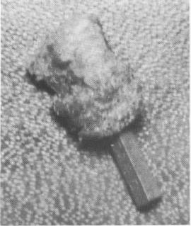
and specimen consists of a metallic pin which is square in outline and 1.2 cm. in length up to the end, where there is a corkscrew-shaped extension. The corkscrew portion is 0.4 cm. in its greatest width, while the square portion is 0.2 cm. in its greatest thickness. Attached to the square portion and apparently fairly loose is a segment of irregular, gray-tan soft tissue which is approximately 0.8 cm. in length and 0.6 cm. in width. The thickest portion extending from the metallic pin is 0.3 cm. The soft tissue is slipped from the metallic pin, and on the pin are the initials Dr. R. C. The metallic pin is placed in storage. The tissue is bi-
Fig. 4-30. The tuberosity implant enclosed by bone.
sected longitudinally, and the cut surfaces are embedded for sectioning. The soft tissue is labeled Specimen A and the calcified tissue labeled Specimen B. Gross photographs and x-rays have been taken.
MICROSCOPIC: A: Sections taken on the implant that had a large radiolucent area encapsulating it reveal irregular segments of active fibrous tissue with small segments of epithelium present. The epithelium in most areas is of a crevicular type, and is supported by a slightly in-flamed granulation tissue. In some of the segments, however, the epithelium is of a more mature gingival type with a small amount of parakeratosis and hyperkeratosis on the surface. Along one margin there is some coagulation of the tissue suggestive of cautery. Because of the folding and irregularity of the fibrous tissue, orientation of the specimen is difficult. However, there appears to be a fairly deep crevicular type epithelial extension along one margin. Some of the collagen bundles are relatively dense and quite irregular.
B: Sections of the decalcified specimen reveal a fairly normal trabeculated bone with consider-able amount of fatty marrow tissue. There are some very prominent cement lines and reversal lines. Normal haversian systems have been distorted by the irregular growth pattern and by some apparent resorption with concomitant re-pair. There are numerous small fragments of
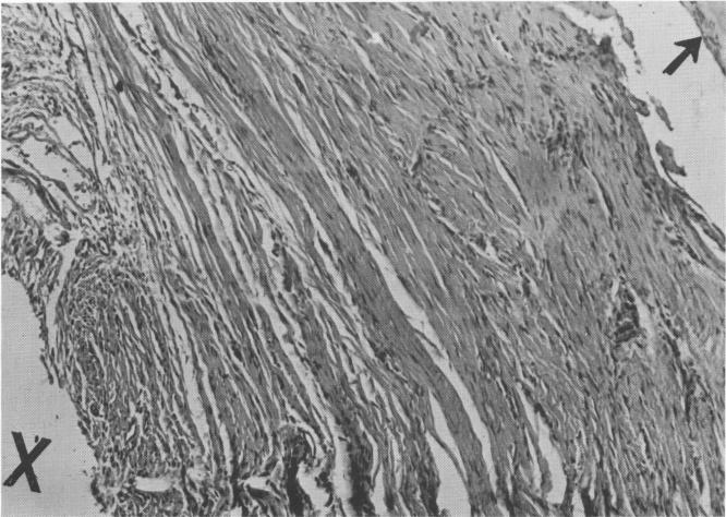
Fig. 4-31. The connective tissue around the successful implant was characterized by its density and regular organization (cross section; high power). X, Implant site; arrow, bone formation.
|
|
Page 98 |
Next Page |
|
Copyright warning: This information is presented here for free for anyone to study online. We own exclusive internet copyrights on all content presented on this website. We use sophisticated technology to identify and legally close down websites that reproduce copyrighted content without permission - so please don’t do it.
|