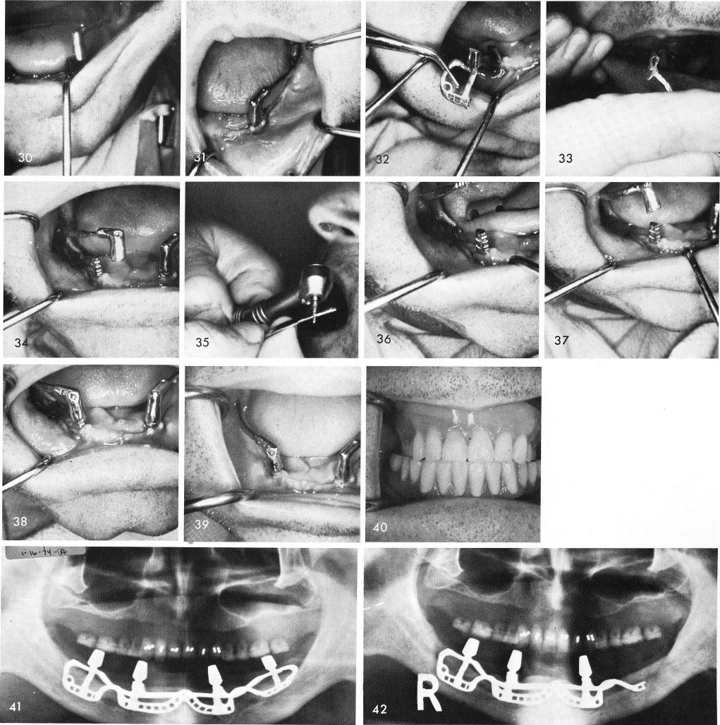
cable and the anterior coping was tapped downward to engage passively the left anterior post, figs. 30, 31. The right posterior quadrant of the subperiosteal implant was then severed and re-moved from the anterior portion, fig. 32, and procedures were duplicated for insertion of the right ramus implant, figs. 33, 34, 35, 36, 37, 38. The healed tissues, fig. 39, and completed denture and x-ray, figs. 40, 41, 42, 43. Fig. 44 shows a failing subperiosteal implant. The two posterior quadrants were failing and had to be severed and removed, figs. 45-46, and sutured, fig. 47. Impressions were taken of the remaining anterior posts for fabrication of the anterior armamentarium, fig. 48. The healing of the tissues around the bilateral sliding cables was excel-