| Mandibular Implants (published 1977) | Dr. Leonard I. Linkow |
|
|
Next Page |
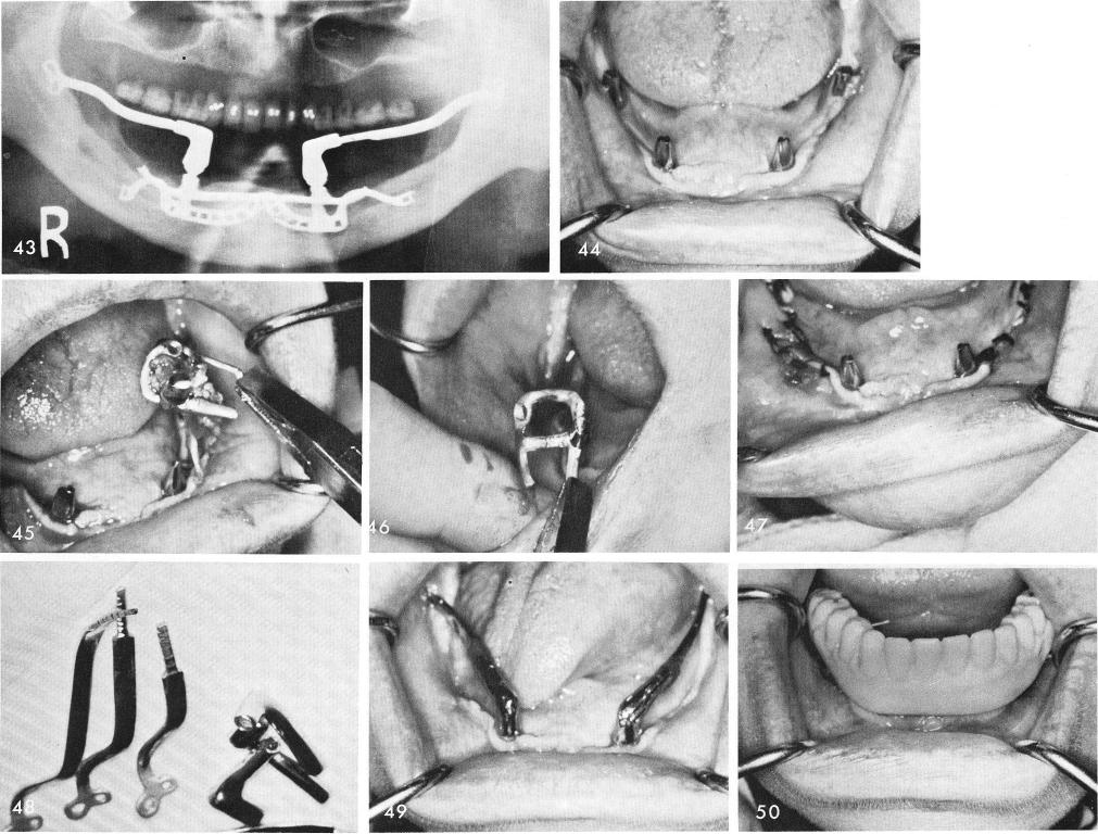
lent, fig. 49. The patient had been wearing this cold cured temporary acrylic splint immediately after surgery, fig. 50. The final denture is locked over the ramus implant with Duralay, fig. 51, and the final occlusion is adjusted, fig. 52. The post-operative x-ray, figs. 53, 54.
The final case which also solved the problem when both posterior quadrants of a subperiosteal implant had failed and was replaced with two ramus sliding cable implants, is seen in fig. 55. A removable denture with horizontal passive attachments (Lew) were very helpful, figs. 56, 57, 58, 59, 60, 61, 62, 63. Fig. 64 shows the final x-ray.
332
|
|
Page 332 |
Next Page |
|
Copyright warning: This information is presented here for free for anyone to study online. We own exclusive internet copyrights on all content presented on this website. We use sophisticated technology to identify and legally close down websites that reproduce copyrighted content without permission - so please don’t do it.
|