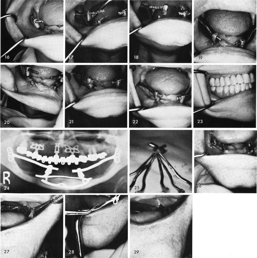
16, 17, 18, 19. They were then tapped upward for cementation, figs. 20, 21, and the posterior tissues were sutured. Healing was excellent, fig. 22, and the full denture was completed and x-rayed, figs. 23, 24. Fig. 25 shows the anterior copings with the horizontal tube extensions that must be prefabricated prior to inserting the ramus implants. Upon clinical observation the left posterior quadrant of the subperiosteal implant was previously removed, fig. 26. The tissues in the left ramus were incised and reflected to expose the bone for making of the ramus groove, fig. 27, and the implant was tapped into correct position, figs. 28, 29. The hollow tube was inserted over the