| Mandibular Implants (published 1977) | Dr. Leonard I. Linkow |
|
|
Next Page |
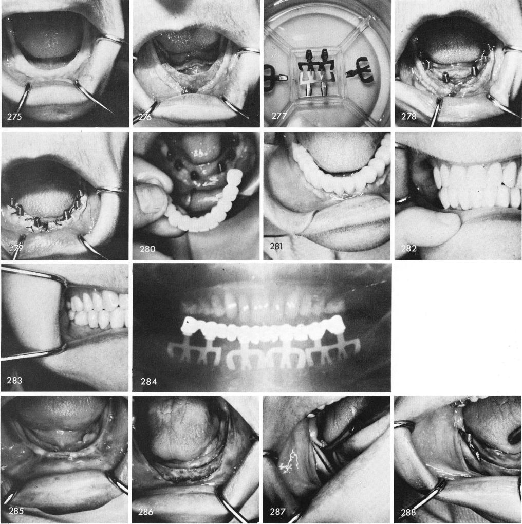
Fig. 275 shows a clinical view of the tissues covering an extremely knife-edge ridge, fig. 276. These early designed blades were used in fig. 277, and tapped properly into the already widened ridge which was accomplished by flattening out the most superior surfaces several milli-meters downward, fig. 278. After suturing, fig. 279, healing was excellent, fig. 280, and the porcelain restoration was cemented into place, figs. 281, 282, 283. The post-operative x-ray, fig. 284.
The only implant in the entire world that can be adapted into knife-edge ridges which is more often the common rather than the uncommon situation that exists are the bladevents. Figs.
242
|
|
Page 242 |
Next Page |
|
Copyright warning: This information is presented here for free for anyone to study online. We own exclusive internet copyrights on all content presented on this website. We use sophisticated technology to identify and legally close down websites that reproduce copyrighted content without permission - so please don’t do it.
|