| Mandibular Implants (published 1977) | Dr. Leonard I. Linkow |
|
|
Next Page |
slip beneath the thickened tissue with the supra-gingival portion of the restorations, flaring out labially and slightly labial to the center of the labial and buccal free marginal gingivae, fig. 261. The bridge and implants remain in place after eight years, fig. 262. The post-operative x-ray, fig. 263.
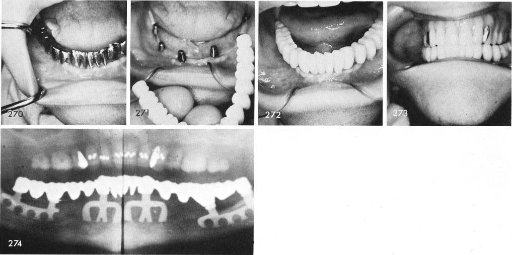
The patient presented herself with a lower denture which she was totally frustrated with, fig. 264. The ridge posteriorly and anteriorly was knife-edged and very shallow, figs. 265, 266. She was warned of a very good chance of a parethesia but she insisted that she did not care and would still rather have the implants, fig. 267. Posteriorly the blades had to go buccally. The tissues were sutured closed, fig. 268, and an immediate temporary acrylic splint was fabricated, fig. 269. Several more visits took place which included the fitting of the gold castings and the completed porcelain-fused to metal prosthesis was ready to be cemented, figs. 270, 271, 272, 273. The x-ray shows the well inserted bladevents. fig. 274.
241
|
|
Page 241 |
Next Page |
|
Copyright warning: This information is presented here for free for anyone to study online. We own exclusive internet copyrights on all content presented on this website. We use sophisticated technology to identify and legally close down websites that reproduce copyrighted content without permission - so please don’t do it.
|