| Theories and Techniques of Oral Implantology (vol.2) (published 1970) | Dr. Leonard I. Linkow |
|
|
Next Page |
| This is an archival HTML version of this book originally hosted here in 2006. The HTML may not display well on modern browsers. Please view the modern PDF Version for a better viewing experience. |
656 Theories and techniques of oral implantology
for a fixed partial denture. A buccal tube was processed to the buccal surface of the crown covering the implant (Fig. 15-92).
The intermaxillary rubber band extended from the maxillary hook attached to the labial arch wire back to the buccal tube on the mandibular crown covering the implant (Fig. 15-93) . It was worn 24
hours a day, thus speeding the reduction of the over-bite and overjet.
This case might also have been treated in the following manner. The implant could have been stabilized to the nearest natural tooth by banding both the tooth and the implant post and extending a .040 rigid wire between them, soldering it to the
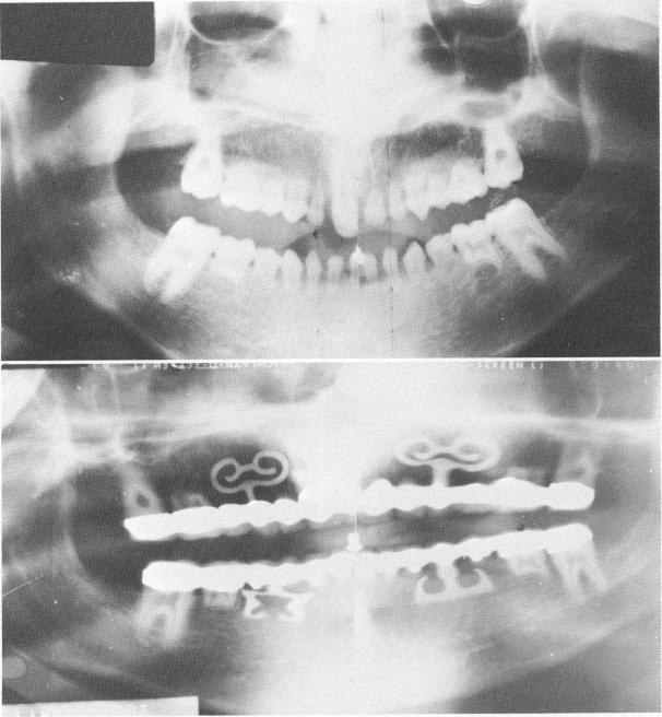
A
B
Fig. 15-89. A, A preoperative Panorex shows the only five permanent teeth. All others were deciduous. B, The Panorex of the completed case, showing the blades.
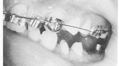
Fig. 15-90. A lower edentulous posterior area with an upper arch wire and molar band with buccal tube is seen.
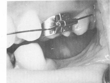
Fig. 15-91. The blade implant post is seen protruding through the fibromucosal tissue. The two bicuspid teeth were prepared for full crown restorations.
|
|
Page 656 |
Next Page |
|
Copyright warning: This information is presented here for free for anyone to study online. We own exclusive internet copyrights on all content presented on this website. We use sophisticated technology to identify and legally close down websites that reproduce copyrighted content without permission - so please don’t do it.
|