| Theories and Techniques of Oral Implantology (vol.2) (published 1970) | Dr. Leonard I. Linkow |
|
|
Next Page |
| This is an archival HTML version of this book originally hosted here in 2006. The HTML may not display well on modern browsers. Please view the modern PDF Version for a better viewing experience. |
Atypical implant situations 653
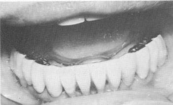
Fig. 15-81. The lower prosthesis was also cemented.
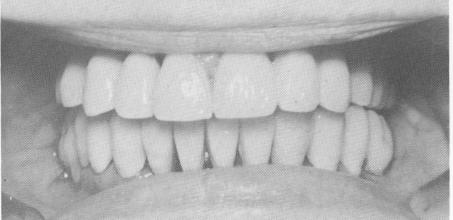
Fig. 15-82. The anterior view of the completed case.
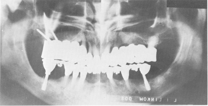
Fig. 15-83. A Panorex showing the pins extending into the zygomatic arch.
patient's face (Fig. 15-75). The left superstructure also included a pink acrylic buccal flange. The pros-thesis was tried in the mouth.
The exact location of the zygomatic arch was determined. (In this case, study skulls and x-rays were used as determinants. Today, however, reflecting the soft tissues to expose the bone is the preferable approach.) Upon determination of the zygomatic arch, holes were made through the template in the direction the pins were to be driven. Then the prosthesis was cemented over the teeth with oxyphosphate of zinc cement (Fig. 15-76). Pin implants were slowly driven through the template in an acute buccal direction in order to enter the zygomatic arch (Fig. 15-77). During pin insertion, x-rays were taken at various intervals to ensure accurate placement.
The superstructures were tried in the mouth (Fig. 15-78) and articulated. Both superstructures were then removed. The pins were fused together with acrylic, and the superstructures were reinserted and cemented (Fig. 1.5-79) .
Endosseous post implants were set into the man-
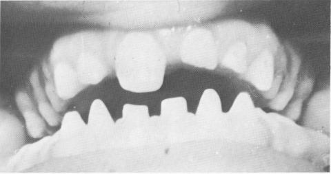
Fig. 15-84. A case of almost complete anodontia of the permanent teeth.
dible (Fig. 15-80) and the lower prosthesis cemented into position (Fig. 15-81). Final adjustments for proper articulation were accomplished in the mouth (Fig. 15-82) . A Panorex shows the case in the mouth (Fig. 15-83).
To date, 5 years later, the fixed full arch denture has been more than satisfactory. The patient has had no pain or discomfort and is much happier than she was with her former restoration.
|
|
Page 653 |
Next Page |
|
Copyright warning: This information is presented here for free for anyone to study online. We own exclusive internet copyrights on all content presented on this website. We use sophisticated technology to identify and legally close down websites that reproduce copyrighted content without permission - so please don’t do it.
|