| Theories and Techniques of Oral Implantology (vol.2) (published 1970) | Dr. Leonard I. Linkow |
|
|
Next Page |
| This is an archival HTML version of this book originally hosted here in 2006. The HTML may not display well on modern browsers. Please view the modern PDF Version for a better viewing experience. |
652 Theories and techniques of oral implantology
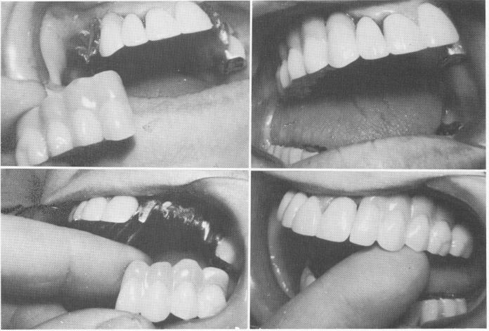
Fig. 15-78. The superstructures were tried.
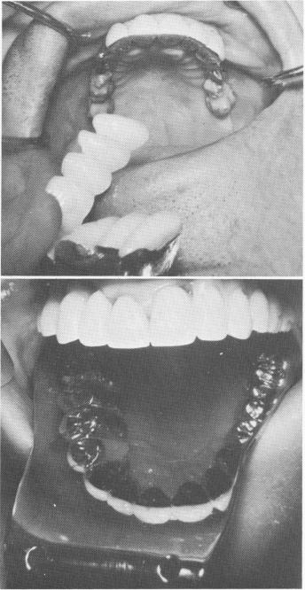
Fig. 15-79. After fusing the pins with acrylic, both super-structures were cemented with hard cement over the templates.
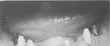
Fig. 15-80. Implant posts were placed in both posterior edentulous areas in the mandible.
ing buccally much further than her opposing re-sorbed and narrower maxillary ridge (Fig. 15-72). Obviously, an unusual approach was necessary.
The teeth were prepared for full crown coverage restorations (Fig. 15-73), and all necessary plaster indices and a wax interocclusal record of centric relation were taken for the completion of a pre-fabricated bridgework. This bridgework consisted of a lower full arch acrylic-and-gold splint, and an upper full arch splint including a bilateral scalloped template with two superstructures (Fig. 15-74) . The template on the right side was built out buccally to contact the mucosal tissue covering the inferior portion of the zygoma. Its superstructure included an acrylic-and-gold veneer crown bridge with a buccal flange of pink resin to extend the right side of the
|
|
Page 652 |
Next Page |
|
Copyright warning: This information is presented here for free for anyone to study online. We own exclusive internet copyrights on all content presented on this website. We use sophisticated technology to identify and legally close down websites that reproduce copyrighted content without permission - so please don’t do it.
|