| Theories and Techniques of Oral Implantology (vol.2) (published 1970) | Dr. Leonard I. Linkow |
|
|
Next Page |
| This is an archival HTML version of this book originally hosted here in 2006. The HTML may not display well on modern browsers. Please view the modern PDF Version for a better viewing experience. |
Subperiosteal implants 567
After the elastic set, the two pins were pulled out of the impression in a labial direction so that the impression could be removed without tearing the material. The two pins were then immediately re-placed into the elastic impression (Fig. 12-89). A surgical wax interocclusal centric relation bite and an alginate impression of the lower arch of teeth were also taken.
A master investment model was poured into the elastic impression with the seated fixation pins (Fig. 12-90), and the subperiosteal framework was waxed up directly on the model. It was then cast after the fixation pins were removed (Fig. 12-91).
After the wound had healed, the tissue was once more anesthetized, incised, and reflected to expose the underlying bone. The sterilized subperiosteal frame-
E
C
B
D
E
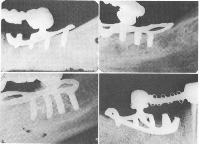
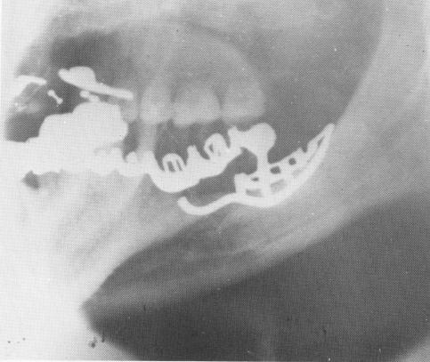
Fig. 12-99. Periapical intraoral x-rays of five different cases utilizing the unilateral subperiosteal implant as the posterior abutments. These cases with the lingual fingers have been in the mouths of these patients from 7 to 12 years. A shows a 12-year postoperative radio-graph.
|
|
Page 567 |
Next Page |
|
Copyright warning: This information is presented here for free for anyone to study online. We own exclusive internet copyrights on all content presented on this website. We use sophisticated technology to identify and legally close down websites that reproduce copyrighted content without permission - so please don’t do it.
|