| Theories and Techniques of Oral Implantology (vol.2) (published 1970) | Dr. Leonard I. Linkow |
|
|
Next Page |
| This is an archival HTML version of this book originally hosted here in 2006. The HTML may not display well on modern browsers. Please view the modern PDF Version for a better viewing experience. |
554 Theories and techniques of oral implantology
the tooth abutments and the implant abutment to immediately stabilize the implant. If a fixation screw is to be used to stabilize the implant, then the acrylic splint is not necessary.
The sutures are removed in about 5 to 7 days. The tissue is usually ready for the final setting of the unilateral implant approximately 3 weeks after the initial bone impression was taken.
To insert the implant, the tissue is incised along the exact line of previous incision, and the soft tissues are gently retracted, exposing the bone. The sterilized implant is then placed into its exact position (Fig. 12-54).
The accuracy of its insertion may be tested by placing the temporary acrylic splint over the implant and natural tooth abutments. If the splint has been fabricated properly and the fit of the implant is ac-curate, there should be no difficulty in positioning the splint. The splint, after being ground into proper occlusion, is then removed from the mouth so that the wound may be closed.
The lips of the wound are closely adapted to the protruding implant post by first making either a mat-tress or a purse-string suture around it and using surgical ties to approximate the remaining tissues.
With a temporary cement, the acrylic splint is set into position to protect the prepared tooth abutments and to immobilize the implant. The splint also places the implant into immediate functional occlusion.
The sutures should be removed anywhere from 7 to 10 days later. If any of the implant frame-work has become exposed, and if this situation was not corrected within 48 hours, there is little sense in resuturing the site. It simply will not heal correctly. The only proper procedure is to remove the implant,
allow the tissue to heal for at least 2 months, and then reincise the tissue and reset and resuture it over the implant a second time. Sometimes in these situations it may be necessary to make new lingual grooves. This, of course, will necessitate taking new impressions for another implant.
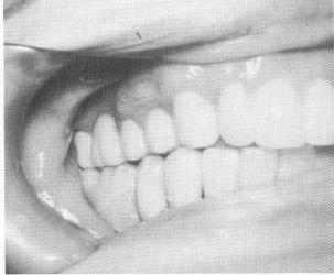
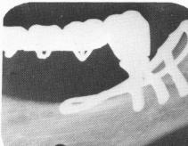
After the tissue has completely healed, the final procedures for completing the fixed partial denture are begun. The previously fitted castings are placed over all the tooth abutments. A gold coping, which was fabricated at the same time as the implant, is fitted over the implant abutment. A wax interocclusal record of centric relation and a plaster index are taken, as well as an alginate impression of the teeth in the opposing jaw for the fabrication of the fixed partial denture. The fixed denture is cemented over the posts using the same procedures as for any ordinary type of fixed partial denture (Fig. 12-55). The x-ray of the completed case is shown in Fig. 12-56.
BILATERAL SUBPERIOSTEAL IMPLANTS
Basically, the surgical procedures for restoring bilaterally edentulous mandibles with partial subperiosteal implants are the same as for unilateral restorations. The remaining teeth are prepared for crown restorations before implant surgery. The first surgical stage for the bilateral bone impression is then done (Fig. 12-57) . A wax bite, plaster index, and opposing upper alginate impression are taken.
If desired—as shown in another very similar case
all of the castings covering the natural tooth abutments and any pontics existing between may be soldered together and the facings processed. However, the casting covering the tooth abutment nearest the implant should be fabricated as a thimble instead of a veneer casting (Figs. 12-58 to 12-60). A veneer
Fig. 12-55. The partial fixed bridge is cemented with hard cement over the implant and tooth abutments.
Fig. 12-56. A periapical postoperative film of the completed case. (From Linkow, L. I.: Re-evaluation of mandibular unilateral subperiosteal implants: a 12 year report, J. Prosth. Dent. 17:509-514, 1967.)
|
|
Page 554 |
Next Page |
|
Copyright warning: This information is presented here for free for anyone to study online. We own exclusive internet copyrights on all content presented on this website. We use sophisticated technology to identify and legally close down websites that reproduce copyrighted content without permission - so please don’t do it.
|