| Theories and Techniques of Oral Implantology (vol.2) (published 1970) | Dr. Leonard I. Linkow |
|
|
Next Page |
| This is an archival HTML version of this book originally hosted here in 2006. The HTML may not display well on modern browsers. Please view the modern PDF Version for a better viewing experience. |
550 Theories and techniques of oral implantology
These partial subperiosteal implants were designed to utilize the same anatomic landmarks as did the full subperiosteal implant. Most other operators also found these implants unsatisfactory because they were unsuitable both in design and method of implantation.
There are basic differences between utilizing a limited site and utilizing the full lower jaw. The lingual tissue covering the mylohyoid ridges is usually much thinner than that in the buccal and occlusal areas. Thus, with the old designs the tissue became extremely taut as it was stretched over the implant. This eventually caused a breakthrough of the lingual border of the implant, with the formation of granulation tissue between the border and the bone, and the implant failed. In those implant designs where
the lingual peripheral border was not extended be-low the mylohyoid ridge, there was very little resistance against lateral forces. Eventual dislodgment of the implant occurred, with granulation tissue growing underneath the implant and dislodging it even more.
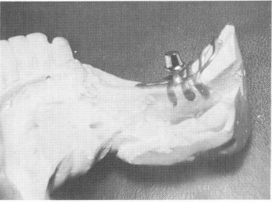
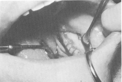
After considering these difficulties, Linkow introduced his open lingual finger unilateral design in 1955. To reduce or eliminate lateral displacement, the lingua] surface of the implant was extended some distance below the mylohyoid ridge, following the contour of the bone (Fig. 12-45). To reduce or eliminate the stretching of the thin lingual tissue over these extensions, notches about 1 mm. deep were made with diamond stones in the bone to accept the fingers (Fig. 12-46). The seated lingual fingers
Fig. 12-45. Lingual fingers (Linkow) extend below the mylohyoid ridge into previously prepared notches to give the unilateral subperiosteal implant added retention and stability. (From Linkow, L. I.: Re-evaluation of mandibular unilateral subperiosteal implants: a 12 year report, J. Prosth. Dent. 17:509-514, 1967.)
A
B
Fig. 12-46. The basal bone is exposed, showing the lingual notches below the mylohyoid ridge. The exposed external oblique ridge is seen on the buccal aspect.
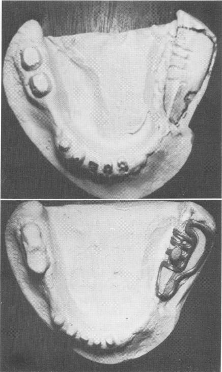
Fig. 12-47. A, The master stone cast illustrating the depth and angulation of the lingual notches as well as the external oblique ridge and part of the mental foramen. B, The implant fits flush onto the stone model and the lingual fingers fit flush into the prepared grooves.
|
|
Page 550 |
Next Page |
|
Copyright warning: This information is presented here for free for anyone to study online. We own exclusive internet copyrights on all content presented on this website. We use sophisticated technology to identify and legally close down websites that reproduce copyrighted content without permission - so please don’t do it.
|