| Theories and Techniques of Oral Implantology (vol.2) (published 1970) | Dr. Leonard I. Linkow |
|
|
Next Page |
| This is an archival HTML version of this book originally hosted here in 2006. The HTML may not display well on modern browsers. Please view the modern PDF Version for a better viewing experience. |
520 Theories and techniques of oral implantology
was waxed, invested, and then cast was made from this modified stone model (Fig. 11-179) .
On the next visit, the tissues were once more incised and reflected to expose the underlying bone. The palatal bar horseshoe blade was then carefully placed into the grooves (Fig. 11-180) and tapped into position (Fig. 11-181) . While tapping the implant, extreme care was taken to guide its insertion so that the palatal portion became flush with the hard palate as the shoulders of the blades became level with the cortical plates of bone.
The tissues were brought snugly together over the shoulders of the implant and around the necks of the abutment posts with surgical ties (Fig. 11-182). About 10 clays later the wound had healed (Fig. 11-183), and the various impression techniques were employed to complete the prosthesis which, in this case, was an all-acrylic splint (Fig. 11-184). It was cemented over the abutment posts with hard cement (Fig. 11-185). A final Panorex shows the ex-tension of the blade across the palate (Fig. 11-186).
The palatal horseshoe type of implant can be considered to be a combined subperiosteal-endosseous implant because of its palatal extension.
Case 17
A palatal horseshoe blade implant for a removable palateless partial denture
The procedures for stabilizing a removable palate-less denture with a horseshoe blade implant are basically similar to those for a fixed denture, except for the final impressions for the prosthesis itself. A re-movable denture rather than a fixed prosthesis be-comes apparent when a sufficient amount of labial bone resorption has taken place to prevent proper esthetics to be attained with a fixed appliance.
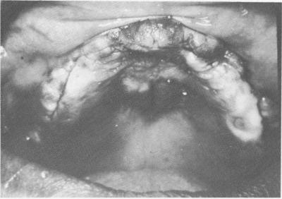
Fig. 11-187. The incision was made along the fibromucosal tissue covering the edentulous maxillary ridge.
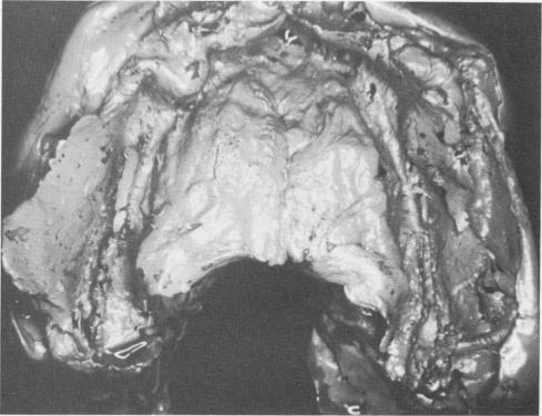
The incision was made from maxillary tuberosity to maxillary tuberosity (Fig. 11-187). The tissues were retracted with a periosteal elevator to expose the entire hard palate. Grooves were then made in the bone and a rubber base impression taken with a specially designed tray. Some impression material was first loaded on the tray while still in a sticky form, while the rest was continously spatulated until it be-came putty-like. This portion was taken in the hands, forced into the shallow grooves, and molded over the ridges and palatal portion ; the loaded tray was immediately placed over it and held in position for about 10 minutes. When it hardened, it was removed (Fig. 11-188), and a bone bite was then taken with wax.
Before the tissues were sutured closed, the thickness of the mucoperiosteum in all areas intended for posts was measured. This was done so that the necks of the implant would not be made too long or too short. The tissues were then sutured together.
The master stone model was poured and articulated with the lower case. The original shallow grooves made in the bone and transferred to the stone model were then deepened according to the radiographic interpretations (Fig. 11-189). The thickness or degree of taper of these grooves in the master stone cast did not matter, since it was intended that the implant be trimmed and tapered to form the characteristic wedge after casting (Fig. 11-190).
At the next visit the tissues were again incised and reflected to insert the implant (Fig. 11-191). The tissues were sutured over the implant (Fig. 11-192). When the tissues had nearly healed (Fig. 11-193),
Fig. 11-188. An elastic impression was made including ex-posed palate and the posterior grooves.
|
|
Page 520 |
Next Page |
|
Copyright warning: This information is presented here for free for anyone to study online. We own exclusive internet copyrights on all content presented on this website. We use sophisticated technology to identify and legally close down websites that reproduce copyrighted content without permission - so please don’t do it.
|