| Theories and Techniques of Oral Implantology (vol.2) (published 1970) | Dr. Leonard I. Linkow |
|
|
Next Page |
| This is an archival HTML version of this book originally hosted here in 2006. The HTML may not display well on modern browsers. Please view the modern PDF Version for a better viewing experience. |
Endosseous blade implants 519
socket, and on the right side, extending as far anteriorly as the buccolingual thickness of the bone permitted (Fig. 11-178). Anteriorly it was impossible to make a groove. Not only was the bone knife-edged, but the entire maxillary process flared out, making it impossible to parallel an anterior groove with the two posterior ones.
The elastic impression included a good portion of the palate as well as the alveolar grooves. From this impression a stone cast was poured. The design and depth of the blades were once again determined by x-rays, and the grooves were made directly in the stone model to coincide with the x-ray findings. A duplicate model on which the horseshoe palatal blade
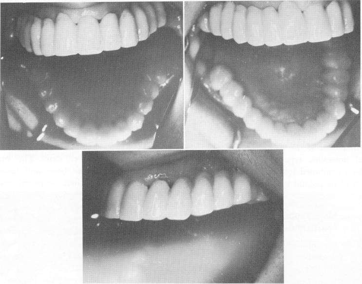
Fig. 11-185. The splint cemented in position.
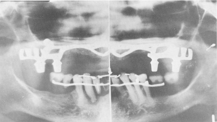
Fig. 11-186. The final Panorex. The mandibular arch was later restored.
|
|
Page 519 |
Next Page |
|
Copyright warning: This information is presented here for free for anyone to study online. We own exclusive internet copyrights on all content presented on this website. We use sophisticated technology to identify and legally close down websites that reproduce copyrighted content without permission - so please don’t do it.
|