| Theories and Techniques of Oral Implantology (vol.2) (published 1970) | Dr. Leonard I. Linkow |
|
|
Next Page |
| This is an archival HTML version of this book originally hosted here in 2006. The HTML may not display well on modern browsers. Please view the modern PDF Version for a better viewing experience. |
484 Theories and techniques of oral implantology
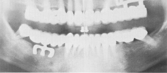
Fig. 11-67. The final Panorex of the completed case.
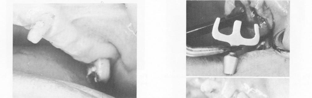
Fig. 11-68. The edentulous span between cuspid and second molar.
After the prostheses were worn for about 5 days, they were removed and the tissues checked for any pontic impingement. All necessary adjustments were made. The teeth and the two full arch prostheses were then thoroughly cleansed and dried before final cementation. A final check was made of the occlusion (Fig. 11-66), and a Panorex of the entire jaw was taken (Fig. 11-67).
Case 6
A full arch splint for a maxilla with long edentulous spans
Only four of this patient's teeth remained: two maxillary cuspids and two molars. For additional support of a full arch restoration, a blade was diagnosed to be placed between the left cuspid and second molar (Fig. 11-68). The tissue was incised and
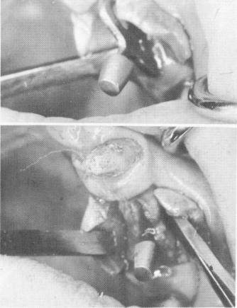
Fig. 11-69. The implant being seated in its site. (From Linkow, L. I.: The blade vent, a new dimension in endosseous implantology, Dent. Concepts 11:3-18, 1968.)
|
|
Page 484 |
Next Page |
|
Copyright warning: This information is presented here for free for anyone to study online. We own exclusive internet copyrights on all content presented on this website. We use sophisticated technology to identify and legally close down websites that reproduce copyrighted content without permission - so please don’t do it.
|