| Theories and Techniques of Oral Implantology (vol.2) (published 1970) | Dr. Leonard I. Linkow |
|
|
Next Page |
| This is an archival HTML version of this book originally hosted here in 2006. The HTML may not display well on modern browsers. Please view the modern PDF Version for a better viewing experience. |
Endosseous blade implants 483
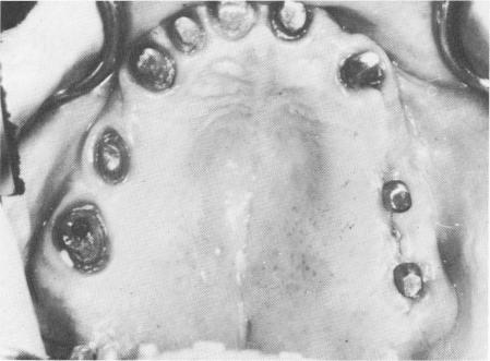
side of the mandible between the cuspid and the wisdom tooth (Fig. 11-62), and the site was closed with simple surgical ties (Fig. 11-63) .
Less than 1 week later the tissue around the implants was almost entirely healed (Fig. 11-64). Both upper and lower porcelain-fused-to-metal full arch dentures were temporarily inserted so that the patient could test them for pain resulting from pontic impingements on the soft tissues. This is very important, since sometimes the pain experienced from impingement is far worse than any pain produced by a faulty implant.
Fig. 11-64. The implant site 5 days later. Considerable healing has taken place.
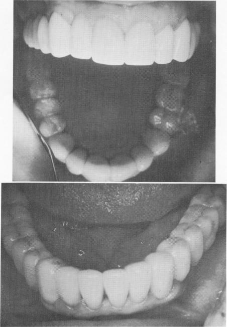
Fig. 11-65. Upper and lower restorations cemented in place.
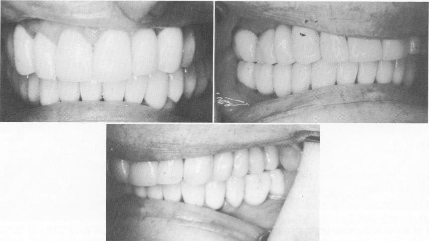
Fig. 11-66. The anterior, left, and right lateral views of the completed case.
|
|
Page 483 |
Next Page |
|
Copyright warning: This information is presented here for free for anyone to study online. We own exclusive internet copyrights on all content presented on this website. We use sophisticated technology to identify and legally close down websites that reproduce copyrighted content without permission - so please don’t do it.
|