| Theories and Techniques of Oral Implantology (vol.2) (published 1970) | Dr. Leonard I. Linkow |
|
|
Next Page |
| This is an archival HTML version of this book originally hosted here in 2006. The HTML may not display well on modern browsers. Please view the modern PDF Version for a better viewing experience. |
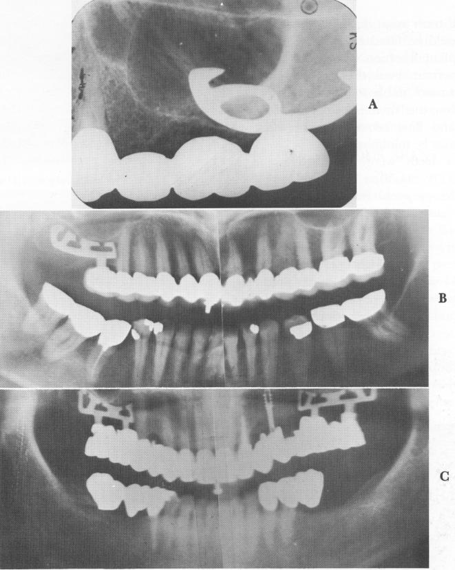
Fig. 11-49. A, A postoperative radiograph reveals the anterior portion in the maxillary sinus. Because no complications occurred, the implant probably rests below the schneiderian membrane. (From Linkow, L. I., and Weiss, J. L.: The endosseous blade: a progress report, Prom. Dent., No. 5, 1969.) B and C, Clear view of how the blades are used to avoid the maxillary sinuses.
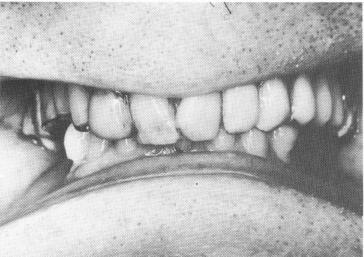
Fig. 11-50. Malpositioned and poorly occluded teeth.
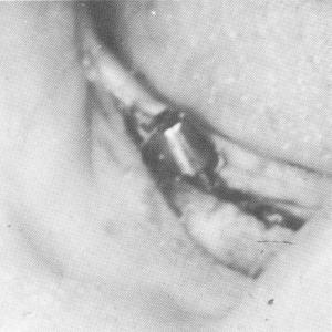
Fig. 11-51. The right mandibular implant in place.
|
|
Page 479 |
Next Page |
|
Copyright warning: This information is presented here for free for anyone to study online. We own exclusive internet copyrights on all content presented on this website. We use sophisticated technology to identify and legally close down websites that reproduce copyrighted content without permission - so please don’t do it.
|