| Theories and Techniques of Oral Implantology (vol.2) (published 1970) | Dr. Leonard I. Linkow |
|
|
Next Page |
| This is an archival HTML version of this book originally hosted here in 2006. The HTML may not display well on modern browsers. Please view the modern PDF Version for a better viewing experience. |
436 Theories and techniques of oral implantology
as was done in this case. The gold copings were placed over the implant shafts and the bridge placed over them. Larger hollows were made in the pontics to avoid impingement. Cold cure acrylic was placed inside the hollowed pontics and the bridge set into position, allowing the acrylic to set inside the mouth. The bridge was removed with the gold copings attached to its tissue-bearing surface. All excess acrylic was trimmed and polished, and the bridge was cemented in place with oxyphosphate of zinc cement.
The triplant pins were then drilled through holes previously made in the bilateral posterior templates (Fig. 10-184). The pins were fastened together with a minimum amount of acrylic to avoid impingement from the superstructures, which were cemented with a loose mix of acrylic (Fig. 10-185). The finished prosthesis was thus supported by the two teeth, four vent-plants, and the two triplants (Fig. 10-186). A Panorex radiograph clearly shows the implant's placement (Fig. 10-187).
Case 20
A full arch splint for a partially edentulous maxilla using internally threaded vent-plants with a screw-on superstructure
Sometimes a screw-on type of fixed partial denture may be preferred instead of the routine type of cemented bridge. If such a prosthesis is desired, it is essential to protect remaining natural teeth against future decay with some form of gold copings. These copings, soldered together, will act as a mesostructure, or internal framework, to which the super-structure can be screwed. The screws holding the superstructure to the mesostructure may be placed through the occlusal surfaces of the superstructure or obliquely angled through the superstructure, as in the following case.
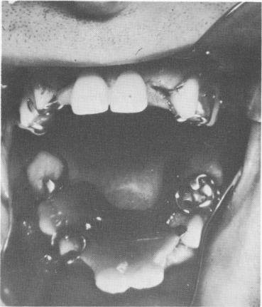
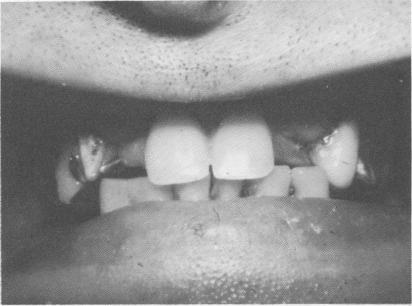
A 52-year-old man with an extremely powerful bite had broken a few of the anterior maxillary teeth off his removable partial denture (Fig. 10-188). His remaining teeth showed rampant caries (Fig. 10-189).
Because his cuspid teeth were so far apart, two internally threaded vent-plants were advisable for the central incisor region.
The remaining teeth in the maxillary arch were prepared for full crown restorations, and all necessary plaster indices and a wax interocclusal record of centric relation were taken. From these, acrylic transfer copings were made and fitted in the mouth (Fig. 10-190). These were picked up with a full mouth plaster index (Fig. 10-191), and from pre-
Fig. 10-188. A broken removable partial denture with two lateral incisor teeth missing from it is all that the patient had been wearing.
Fig. 10-189. The poor condition of the remaining teeth is clearly evident.
Fig. 10-190. The teeth were prepared for full crown restorations, and acrylic transfer copings were fitted over them.

|
|
Page 436 |
Next Page |
|
Copyright warning: This information is presented here for free for anyone to study online. We own exclusive internet copyrights on all content presented on this website. We use sophisticated technology to identify and legally close down websites that reproduce copyrighted content without permission - so please don’t do it.
|