| Theories and Techniques of Oral Implantology (vol.2) (published 1970) | Dr. Leonard I. Linkow |
|
|
Next Page |
| This is an archival HTML version of this book originally hosted here in 2006. The HTML may not display well on modern browsers. Please view the modern PDF Version for a better viewing experience. |
Maxillary endosseous implant interventions 433
ample, in order to avoid a low-flaring sinus floor, a pin may have to be so acutely angled that its protruding end may excessively extend along the template. It thus becomes necessary to make two acrylic cores instead of one in order to follow the contours of the template. In this manner the pins are still locked to one another to form a tripod, even though
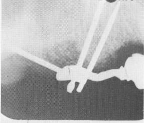
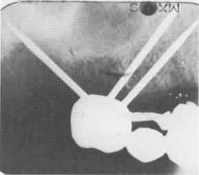
Fig. 10-179. Roentgenograms show the pins drilled through the template and the superstructure cemented over the pins.
one pin may be inside one core while the others are inside the second core. As long as these two cores are joined interproximally, the pins are secure.
When the cores hardened, they were prepared for full coverage restorations (Fig. 10-177). The superstructure was then fitted, adjusted, and cemented into position over the two acrylic cores and gold coping (Fig. 10-178). Fig. 10-179 shows the pins driven through the scalloped template and the superstructure cemented over the acrylic core and gold coping.
Case 19
A full arch prefabricated splint for a partially edentulous maxilla using vent-plants and bilateral triplants
One of the more practical applications of pre-fabricated splints is that the patient's own dentist can work him up to a point where the implantologist need only insert the implants. Thus a dentist inexperienced in the insertion of implants can do the bridgework construction and follow up, radiographically and clinically, the progress of the implants placed in his patient's mouth. This approach was used successfully on a 53-year-old male patient. His dentist, with guidance from Linkow as to design and articulation, made the prosthesis. The implants were then inserted and adapted to the bridge.
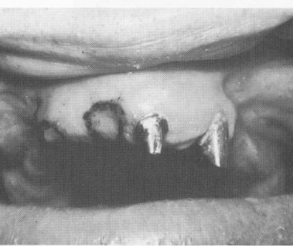
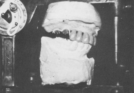
The patient only had two remaining teeth in the maxillary arch, a left central and left cuspid. These were prepared and covered with gold copings by his regular dentist (Fig. 10-180). A prefabricated full arch fixed denture was processed on a Hanau articulator (Fig. 10-181) .
Fig. 10-180. Only two teeth remained in the entire arch.
Fig. 10-181. The prefabricated full arch fixed denture was fabricated with the aid of a Hanau articulator.
|
|
Page 433 |
Next Page |
|
Copyright warning: This information is presented here for free for anyone to study online. We own exclusive internet copyrights on all content presented on this website. We use sophisticated technology to identify and legally close down websites that reproduce copyrighted content without permission - so please don’t do it.
|