| Theories and Techniques of Oral Implantology (vol.1) (published 1970) | Dr. Leonard I. Linkow |
|
|
Next Page |
| This is an archival HTML version of this book originally hosted here in 2006. The HTML may not display well on modern browsers. Please view the modern PDF Version for a better viewing experience. |
116 Theories and techniques of oral implantology
Studies of bone blocks including crystalline bone screws consistently revealed the absence of connective tissue between the implant and the bone (Fig. 4-72). The bone apparently grows right up to crystalline implants. This is totally unlike the typical histologic picture of metallic implants, and further investigations are being conducted to investigate the significance of these differences.
In summary, Blakey concluded that both metallic and crystalline implants are accepted. Very little osteoclastic activity was observed. There were also rare findings of inflammatory cells. All metallic implants were separated from the bone by a connective tissue membrane; crystalline implants were not.
Hodosh, Povar, and Shklar on plastic implants
Hodosh, Povar, and Shklar reported in the Journal of the American Dental Association (1965)
lllr'
their histologic findings on plastic implants in dogs, Macaca irus monkeys, Rhesus monkeys, and baboons.
As implants they used exact replicas of extracted teeth. These were made of methyl methacrylate and were fashioned immediately after tooth extraction. Thirty minutes after tooth removal the site was occupied by the plastic replica. The implant was Immediately fixed to adjacent teeth either with stainless steel wire and acrylic or by an intrabony horizontal pin technique, driving a Vitallium pin from the buccal plate of bone through the plastic root and ending in the lingual plate of bone.
Some of the implants had holes through the apical portions to encourage bony bridging (Fig. 4-73), others did not. Interestingly enough, when the holes were made near the crown, a foci of infection associated with gingival recession occurred. To avoid this, the holes were positioned more apically
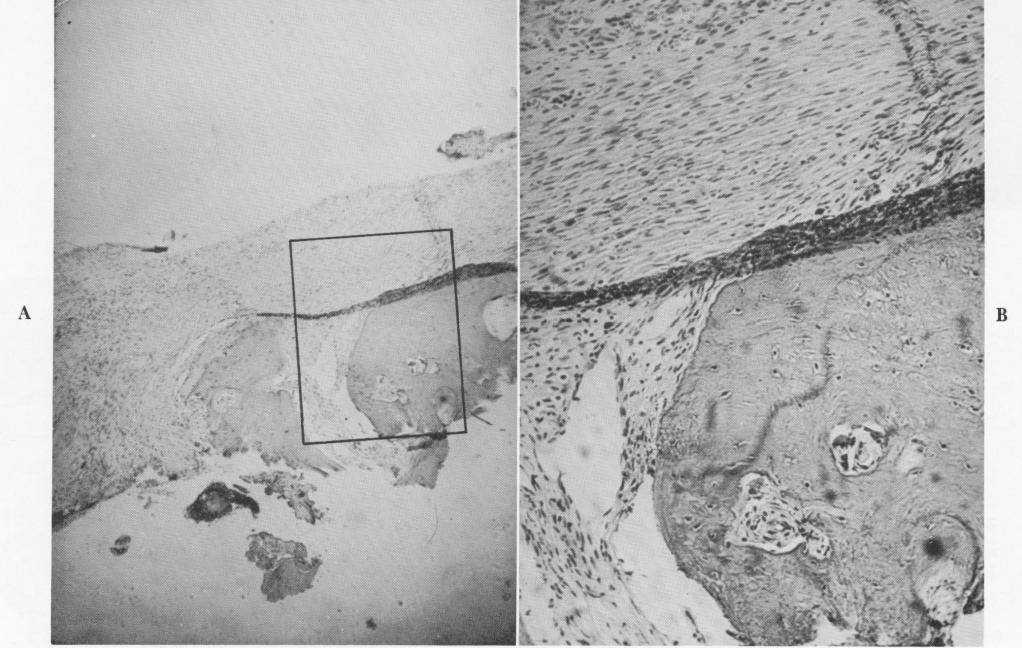
Fig. 4-71. A, Section through the tissue found embedded in the vent of a vent-plant. Under low power both fibrous tissue and bone are evident. B, A high-power view reveals the healthy nature of the bone inside the vent, as evidenced by the osteocytes. (Courtesy K. R. Blakey, Howard University.)
|
|
Page 116 |
Next Page |
|
Copyright warning: This information is presented here for free for anyone to study online. We own exclusive internet copyrights on all content presented on this website. We use sophisticated technology to identify and legally close down websites that reproduce copyrighted content without permission - so please don’t do it.
|