| Theories and Techniques of Oral Implantology (vol.1) (published 1970) | Dr. Leonard I. Linkow |
|
|
Next Page |
| This is an archival HTML version of this book originally hosted here in 2006. The HTML may not display well on modern browsers. Please view the modern PDF Version for a better viewing experience. |
Implant histology 131
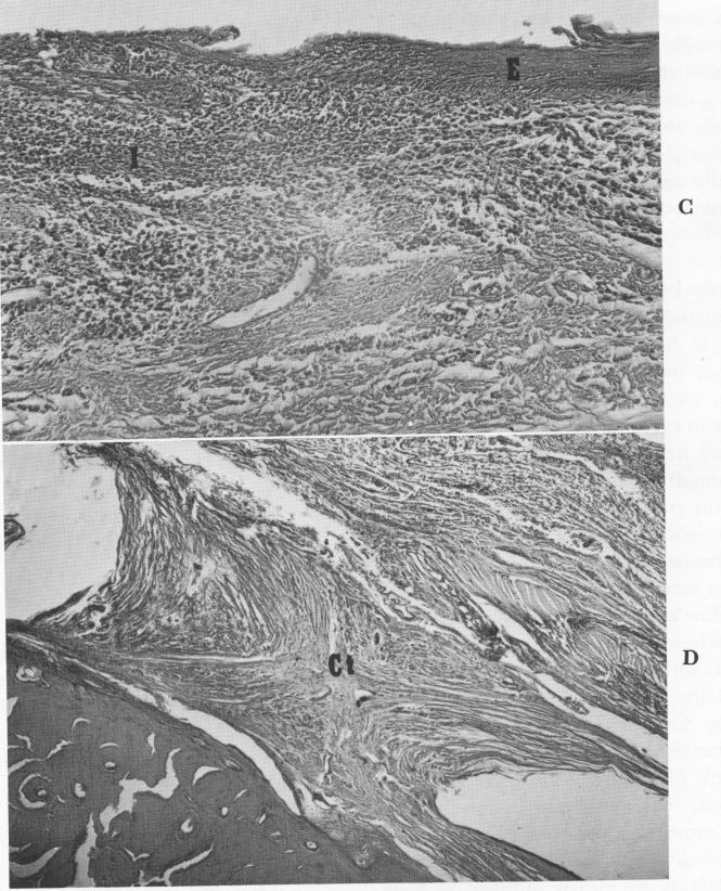
and the bone, was the thickest soft tissue observed in all the sections. An epithelial layer fifteen to twenty cells deep lined its surface, and the underlying connective tissue contained inflammatory cells that in-creased in number on the buccal and lingual sites of the abutment. The epithelium lining the space occupied by the primary strut decreased until it faded out completely 2 to 3 mm. from the penetration site and was replaced by typical implant connective tissue. At this point, the inflammatory cells accompanying the epithelial invagination increased, suggesting that epithelial invagination along a strut is related to inflammation. Considering the small amount of invagination over a 12-year period, the findings are most significant and encouraging.
Bodine and Mohammed concluded that although the epithelium did grow downward along the struts, this invagination did not extend more than a few millimeters away from the site where the posts penetrated the fibromucosa. However, because these posts were sources for epithelial invagination, they suggest limiting the number of abutment posts to four. Also, secondary metal exposures should be avoided.
The connective tissue adjoining the buried metal portions was normal and dense. Those layers immediately facing the metal contained flattened, elongated cells that gave those layers a tendon-like character.
Although this patient had had his implant over 12 years and had strong masticatory habits, there
Fig. 4-103, coned. For legend see opposite page.
|
|
Page 131 |
Next Page |
|
Copyright warning: This information is presented here for free for anyone to study online. We own exclusive internet copyrights on all content presented on this website. We use sophisticated technology to identify and legally close down websites that reproduce copyrighted content without permission - so please don’t do it.
|