| Theories and Techniques of Oral Implantology (vol.1) (published 1970) | Dr. Leonard I. Linkow |
|
|
Next Page |
| This is an archival HTML version of this book originally hosted here in 2006. The HTML may not display well on modern browsers. Please view the modern PDF Version for a better viewing experience. |
Single tooth implants 325
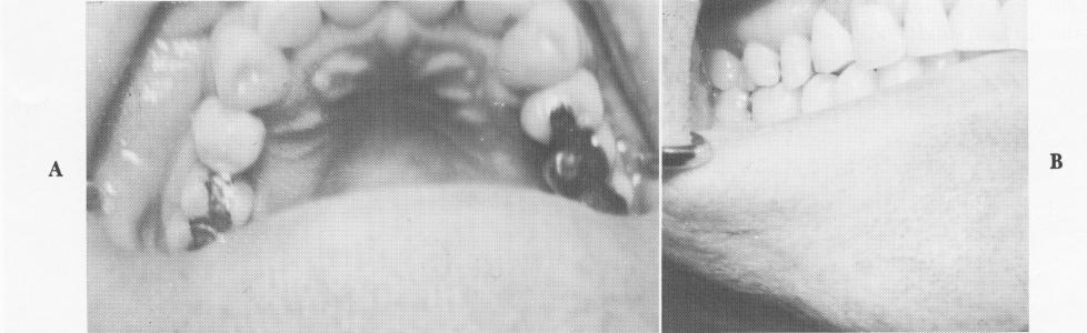
Fig. 8-45. A, The acrylic crown is cemented over implant shaft and the attached inlay cemented into its prepared seat. B, The completed case as viewed from the side.
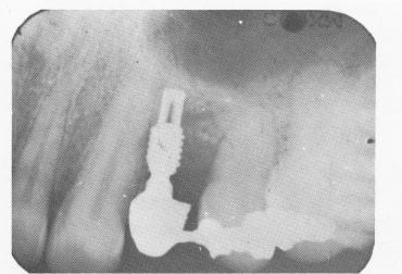
Fig. 8-46. A periapical postoperative radiograph of the completed case.
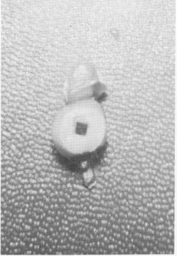
Fig. 8-47. The undersurface of a maxillary molar acrylicand-gold occlusal crown with the square-shaped lumen that fits over the implant shaft. Extending from the crown on its mesial and distal proximal surfaces are two gold inlays that add greatly to the support of the implant.
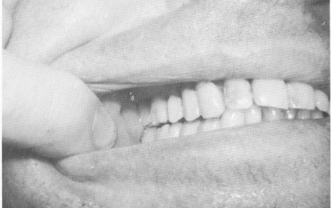
Fig. 8-48. The implant post extending from the fibromucosal tissue in the endentulous area is seen between the bicuspid and molar teeth. Notice the mesiocclusal preparation on the molar.
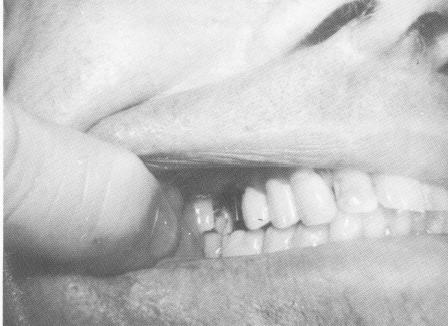
Fig. 8-49. The molar crown is cemented over the implant post, and the inlays extending from both of its interproximal surfaces are cemented into the inlay preparations on both neighboring teeth.
|
|
Page 325 |
Next Page |
|
Copyright warning: This information is presented here for free for anyone to study online. We own exclusive internet copyrights on all content presented on this website. We use sophisticated technology to identify and legally close down websites that reproduce copyrighted content without permission - so please don’t do it.
|