| Theories and Techniques of Oral Implantology (vol.1) (published 1970) | Dr. Leonard I. Linkow |
|
|
Next Page |
| This is an archival HTML version of this book originally hosted here in 2006. The HTML may not display well on modern browsers. Please view the modern PDF Version for a better viewing experience. |
332 Theories and techniques of oral implantology
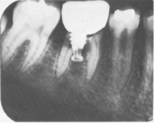
Fig. 8-75. A 4-year postoperative radiograph. Root canal therapy could not be done since the tooth split exactly through the center of the canals. The translucencies seen along both roots are caused by internal resorption.
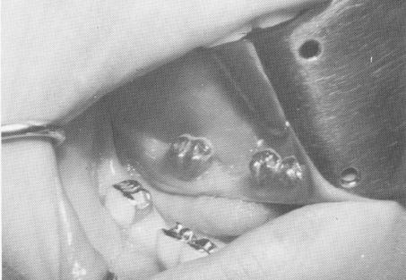
Fig. 8-76. Class II inlay preparations were prepared insid the gold work of the molar crown and second bicuspi crown.
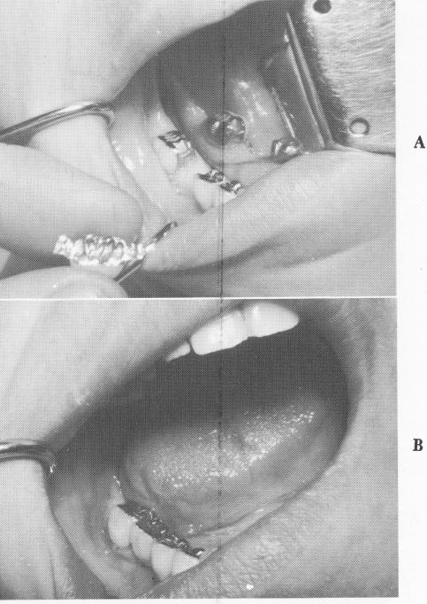
Fig. 8-79. A, The restoration is fitted into position prior to the insertion of the implant. B, The fit of both inlays, the gingival adaptation of the molar crown, and the accuracy of occlusion are carefully checked.
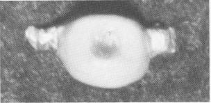
Fig. 8-77. An acrylic veneer molar crown with two inlays attached to it is fabricated.
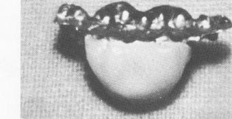
Fig. 8-78. The tissue-bearing surface of the one-piece c ing. The hole in its center fits over implant shaft.
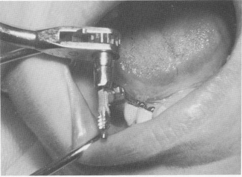
Fig. 8-80. The vent-plant with a Dacron sleeve attached is screwed into the bone.
|
|
Page 332 |
Next Page |
|
Copyright warning: This information is presented here for free for anyone to study online. We own exclusive internet copyrights on all content presented on this website. We use sophisticated technology to identify and legally close down websites that reproduce copyrighted content without permission - so please don’t do it.
|