| Theories and Techniques of Oral Implantology (vol.1) (published 1970) | Dr. Leonard I. Linkow |
|
|
Next Page |
| This is an archival HTML version of this book originally hosted here in 2006. The HTML may not display well on modern browsers. Please view the modern PDF Version for a better viewing experience. |
Single tooth implants 333
graph clearly illustrates the solution (Fig. 8-75). There has been no problem whatsoever to date—4 years later—because the implant is held firmly by the lingual portion of the molar tooth.
Case 10
Replacing a missing pontic in an existing fixed partial denture with a single implant
This is a case where an implant was used to secure and stabilize a four-unit, lower right fixed partial denture made several years previously. The patient, a 41-year-old woman, presented herself with the remains of a fixed partial denture. The solder joints on both sides of the pontic between the last abutment and the second bicuspid crown broke, and the patient lost the pontic.
The mesiocclusal crown of the last molar and the distocclusal crown of the second bicuspid were immediately prepared for inlay preparations (Fig. 8-76). A hydrocolloid impression of both inlays and of the edentulous space was taken, as well as an opposing alginate and wax interocclusal record of centric relation. A molar crown with two interproximal extension inlays was cast and an acrylic facing processed on the buccal surface of the crown (Fig. 8-77). A hole was made on the tissue-bearing surface of the molar crown to accommodate the implant shaft (Fig. 8-78).
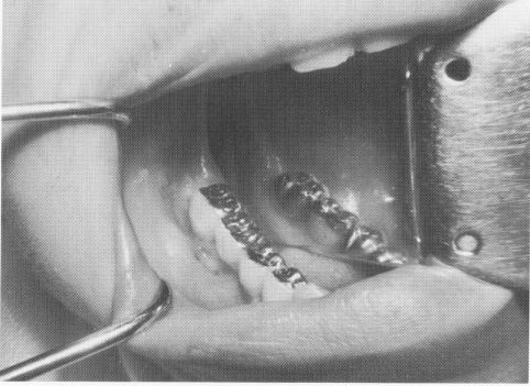
The restoration was tried in the mouth before the implant was inserted, and all necessary adjustments were accomplished (Fig. 8-79). The restoration was then removed, and the vent-plant with a Dacron sleeve was screwed into the bone (Fig. 8-80). (It was hoped that the porous character and slightly irritating quality of the Dacron would pro-mote closer adaptation of the collagenous tissue around the post. Approximately three dozen cases using Dacron sleeves screwed into bone have been done, and all are still in the mouths functioning well. The results are still unsubstantiated by tissue biopsies.) The restoration was then cemented into position with hard cement (Fig. 8-81) and radiographed (Fig. 8-82).
IMMEDIATE SINGLE TOOTH IMPLANTATIONS FOLLOWING AN EXTRACTION
It is very tempting to try a single tooth implantation in an open socket. The idea is appealing to both the dentist and the patient. However, there are numerous difficulties that are often practically insurmountable.
Fig. 8-81. The prosthesis is cemented over the implant post and into the Class II inlay preparations.
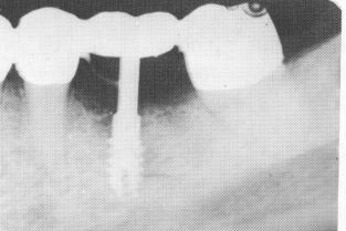
Fig. 8-82. A postoperative radiograph showing the pre-fabricated restoration locked into the existing bridge sup-porting the implant.
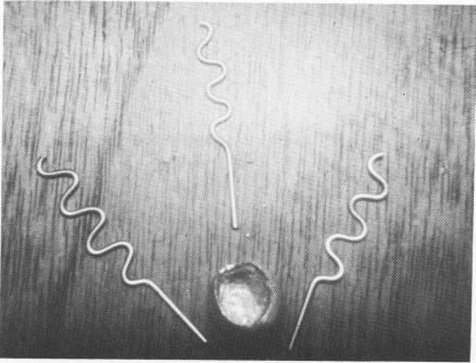
Fig. 8-83. Three twisted pins for insertion into an open socket. A prefabricated acrylic-fused-to-gold thimble crown is also seen.
|
|
Page 333 |
Next Page |
|
Copyright warning: This information is presented here for free for anyone to study online. We own exclusive internet copyrights on all content presented on this website. We use sophisticated technology to identify and legally close down websites that reproduce copyrighted content without permission - so please don’t do it.
|