| Theories and Techniques of Oral Implantology (vol.1) (published 1970) | Dr. Leonard I. Linkow |
|
|
Next Page |
| This is an archival HTML version of this book originally hosted here in 2006. The HTML may not display well on modern browsers. Please view the modern PDF Version for a better viewing experience. |
96 Theories and techniques of oral implantology
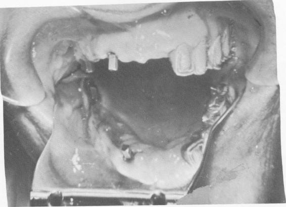
Fig. 4-25. With the impacted tooth left undisturbed, three spiral-shaft implants were inserted.
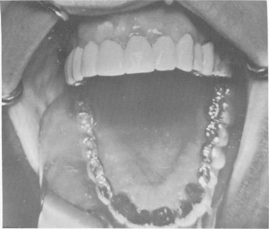
Fig. 4-26. The restoration in place.
be removed first. This unique situation provided another opportunity for obtaining a human bone block specimen.
According to the lateral plate (Fig. 4-28), the two implants in the tuberosity were solid. However, only one was successfully removed with the bone still attached. The implant directly underneath the crown portion of the impacted cuspid was loose and bone loss evident (Fig. 4-29); it was removed with connective tissue attached. The tuberosity implant with bone attached and the failing implant were
submitted for examination (Fig. 4-30). The specimens were sectioned and photomicrographs taken (Fig. 4-31).
The following is the histologic report by Dr. Francis Howell, Jr., of La Jolla, California:
GROSS: The specimens were received in formalin. One is a roughly rectangular segment of bone with a crescent-shaped concavity in one margin. The entire specimen measures 0.7 x 0.6 cm. in area and is 0.35 cm. in its greatest thickness. The greatest radius of the concavity is 0.2 cm. The sec-
|
|
Page 96 |
Next Page |
|
Copyright warning: This information is presented here for free for anyone to study online. We own exclusive internet copyrights on all content presented on this website. We use sophisticated technology to identify and legally close down websites that reproduce copyrighted content without permission - so please don’t do it.
|