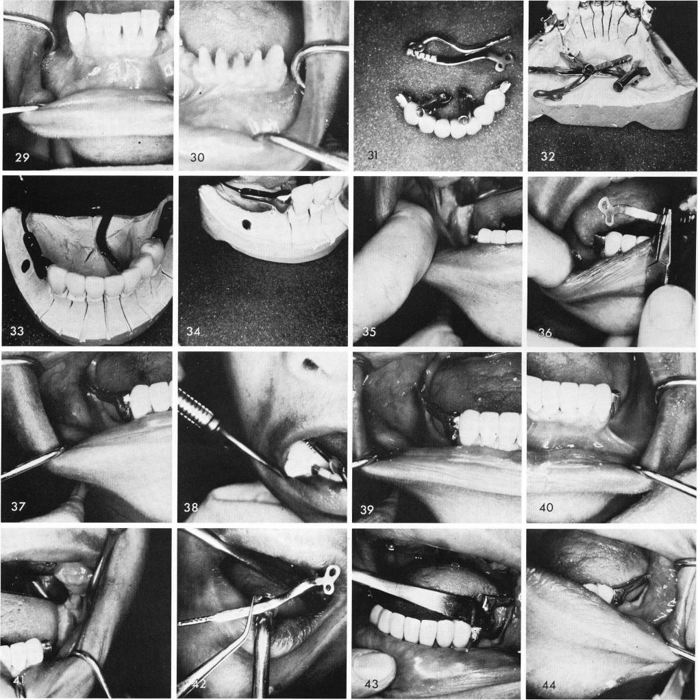
Another case showing remaining anterior teeth, fig. 29, that were prepared for the anterior fixed prosthesis, fig. 30. The anterior prosthesis was fabricated with two cantilevered pontics to accept two telescopic copings with distally extended hollow tubes, figs. 31, 32, 33, 34. The right ramus groove is made, fig. 35, and the ramus blade is inserted, fig. 36, tapped into proper position, fig. 37. It is then tapped upward for disengagement of the parts and final cementation, figs. 38, 39. The same procedure is followed on the other side, figs. 40, 41, 42, 43, 44. The bridge is then completed, figs. 45, 46. The post-operative x-ray, fig. 47.
323