| Mandibular Implants (published 1977) | Dr. Leonard I. Linkow |
|
|
Next Page |
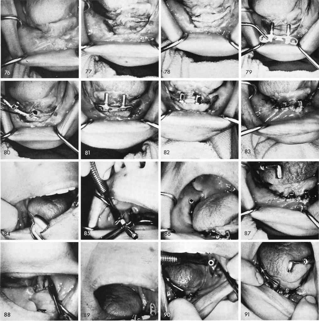
Fig. 76 shows an extremely resorbed mandible with scarred tissues caused by a subperiosteal implant that failed. The bone is exposed, fig. 77, and groove made, fig. 78. The blade is fitted and tapped into position, figs. 79, 80, 81, 82, and the tissues are sutured closed, fig. 83. The grooves are made in both rami and the various parts of the system are inserted and locked together, figs. 84, 85, 86, 87, 88, 89, 90, 91, 92. The parts are then separated so that the acrylic locking medium
308
|
|
Page 308 |
Next Page |
|
Copyright warning: This information is presented here for free for anyone to study online. We own exclusive internet copyrights on all content presented on this website. We use sophisticated technology to identify and legally close down websites that reproduce copyrighted content without permission - so please don’t do it.
|